Download
1 / 58
580 likes | 811 Vues
Postpartum Hemorrhage. Xiong yu Obstetric & Gynecology Hospital , Fudan University. Blood loss in excess of 500 ml following birth within the first 24 hours of delivery Serious intrapartum complication

E N D
Postpartum Hemorrhage Xiong yu Obstetric & Gynecology Hospital, Fudan University
Blood loss in excess of 500 mlfollowing birth within the first 24 hours of delivery Serious intrapartum complication The most significant cause of maternal death worldwide, mortality : 140 000 per year (1 maternal death every 4 minutes) Incidence: 4–6% of pregnancies Actual incidence: more high because of inaccurate, significant underreporting
Primary PPH Occurring within the first 24 hours of delivery 4–6% of pregnancies Caused by uterine atony in 80% or more of cases Secondary PPH Occurring between 24 hours and 6–12 weeks postpartum 1% of pregnancies
4 “ T ” Tone: uterine atony Tissue: retained placenta Trauma: vaginal, cervical, or uterine injury Thrombin: coagulopathy (pre-existing or acquired) ——SOGC guideline (number 235, October 2009): Active Management of the Third Stage of Labor: Prevention and Treatment of Postpartum Hemorrhage
The most common and important cause of PPH The primary protective mechanism for immediate hemostasis after delivery: Myometrial contraction causing occlusion of uterine blood vessels ——living ligatures of the uterus Blood flow from the vascular space to the uterine cavity via the myometrium is impeded
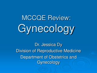
Placenta previa Placenta abruption 胎儿 子宫内膜 出血 脐带 胎盘 宫颈
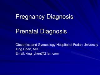
Uterine anomalies Twin pregnancy fibroid 带蒂 内膜下肌瘤 胎盘 肌壁间肌瘤 脐带 脐带 浆膜下肌瘤 内膜下肌瘤 胎儿 胎儿 带蒂 浆膜下肌瘤 宫颈 阴道
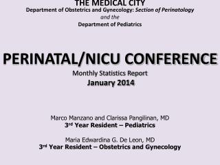
Placenta villi attach Placenta villi invade Placenta villi penetrate to the myometrium into the myometrium through the myometrium Accreta Increta Percreta
II I Laceration of cervix III Lacerations of perineum
Vaginal bleeding Bleeding with characteristic soft, poorly contracted (“boggy”) uterus on bimanual pelvic examination ——uterine atony Bleeding while the uterus is firmly contracted —— retained placenta ——genital tract laceration Bleeding without clot ——coagulopathy Pelvic or rectal pressure and pain ——genital tract hematomas
Hypovolemic shock • Irritable,pallor and clamminess of skin,tachycardia, narrow pulse pressure ——mild degree of shock
Weight method: • Blood loss(ml)=(dressing wet weight after birth-dressing dry weight before birth)/1.05(specific gravity of blood) • Volume method: • Collect blood using a container • Area method: • 10cm*10cm gause soak blood = 10ml blood
Shock index =heart rate/systolic pressure(mmHg) (normal <0.5) shock index estimate loss of blood(ml) loss of blood volume 0.6~0.9 <500~750 <20% =1.0 1000~1500 20~30% =1.5 1500~2500 30~50% ≥2.0 2500~3500 ≥50~70%
The initial goal Identifying and treating the cause of blood loss Instituting resuscitative measures to maintain hemodynamic stability and oxygen perfusion of the tissues
Call for help Resuscitation Assess the “ABC” Monitor BP, P, R Empty bladder, monitor urine output IV line Crystalloid, isotonic fluid replacement Oxygen by mask Laboratory tests Complete blood count Coagulation screen Blood grouping and cross ——SOGC 2009
Uterine massage Diminish bleeding, expel blood and clots, and allow time for other measures to be implemented Uterotonic drugs Ongoing blood loss in the setting of decreased uterine tone requires the administration of additional uterotonics as the first-line treatment for hemorrhage
Uterine tamponade Exploratory laparotomy Uterine artery embolization
Indication:uterotonics fail to cause sustained uterine contractions and satisfactory control of hemorrhage after vaginal delivery
Packing Bakri Balloon tamponade
Indication:When uterotonic agents with or without tamponade measures fail to control bleeding in a patient who has given birth vaginally Techniques Compression sutures Artery ligation Hysterectomy
B-Lynch technique First reported by B-lynch in 1993 Compress the uterine corpus and decrease bleeding Rare Complication:uterine ischemic necrosis with peritonitis
Modified B-Lynch e.g. Hemostatic multiple square suturing For postpartum hemorrhage caused by uterine atony, placenta previa, or placenta accreta Eliminateing space in the uterine cavity by suturing both anterior and posterior uterine walls
Bilateral uterine arteries ligation Bilateral internal iliac arteries ligation Bilateral ovarian arteries ligation
Uterine arteries ligation Internal iliac arteries ligation
Diminish the pulse pressure of blood flowing to the uterus The timing of this intervention is important: it must be done without delay, before excessive blood loss has occurred Surgical skill is required to avoid failure and complications such as damage to other vascular structures and the ureters
Indication: massive hemorrhage has not responded to previous interventions Notice: If hysterectomy is performed for uterine atony, there should be documentation of other therapy attempts
Hysterectomy cavity cavity uterus salpinx endometrium overy myometrium subtotal cervix bladder total vagina
Indication: stable vital signs , persistent bleeding, especially if the rate of loss is not excessive Used for bleeding that continues after hysterectomy Used as an alternative to hysterectomy to preserve fertility
Radiographic identification of bleeding vessels Embolization with gelfoam, coils, or glue, or balloon occlusion
H.A.E.M.O.S.T.A.S.I.S. H: Ask for help A: Assess (vital parameters, blood loss) and resuscitate E: Establish etiology and check medication supply (oxytosin, ergometrine) and availability of blood M: Massage uterus O: Oxytocin infusion, prostaglandins (intravenous, rectal, intramuscular, intra- myometrial)
S: Shift to operating room, exclude retained products and trauma, bimanual compression T: Tamponade balloon, uterine packing A: Apply compression sutures S: Systematic pelvic devascularization (uterine, ovarian, internal iliac) I: Intervention radiologist, uterine artery embolization if appropriate S: Subtotal or total abdominal hysterectomy ——ICM/FIGO guideline 2006: Postpartum hemorrhage today: initiative 2004—2006
Diagnosis: detection of an echogenic mass in the uterus by ultrasonography Directed therapy Whole placenta in uterus:manual removal Incomplete separation (avulsed lobule, succenturiate lobe):gentle curettage Placenta accreta curettage wedge resection medical management hysterectomy
Lacerations of perineum, vagina, or cervix Genital tract hematomas
Identification and proper repair of lacerations Transfer to a well-equipped operating room Proper patient positioning Adequate operative assistance Good lighting Appropriate instrumentation (eg, Simpson or Heaney retractors) Adequate anesthesia
May not be recognized until hours after the delivery Sometimes occur in the absence of vaginal or perineal lacerations The main symptoms are pelvic or rectal pressure and pain
Directed therapy Draining the blood within the hematoma (sometimes placing a drain in situ) Suturing the incision Packing the vagina Interventional radiology
Directed therapy Appropriate testing Blood products infused as indicated Simultaneous surgery if the coagulopathy caused or perpetuated by the hemorrhage