Investigating JIL-1 Kinase's Role in Lamin Interaction and Protein Purification Techniques
10 likes | 137 Vues
This research explores the functional analysis of the JIL-1 kinase and its interactions with lamin, focusing on protein purification methods and X-ray crystallography. We evaluated the lamin migration pattern on SDS-PAGE gels from wild-type and mutant Drosophila, alongside S2 cell extracts. Our objective was to optimize purified protein yields and examine the kinase's potential impact on lamin phosphorylation. Through various purification methods, we aim to understand the structure of JIL-1 and its biological significance in nuclear architecture.

Investigating JIL-1 Kinase's Role in Lamin Interaction and Protein Purification Techniques
E N D
Presentation Transcript
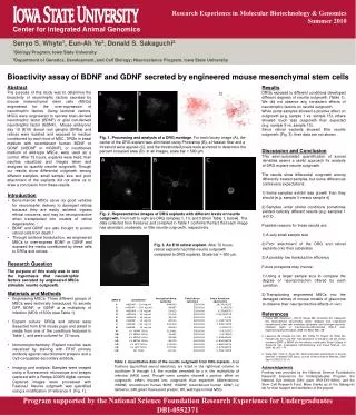
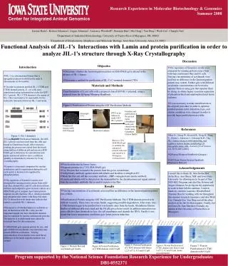
Research Experience in Molecular Biotechnology & Genomics • Summer 2008 Center for Integrated Animal Genomics Jessian Resto1; Kristen Johansen2; Jorgen Johansen2; Laurence Woodruff2; Xiaomin Bao2; Hui Deng2; Yun Ding2; Weili Cai2; Changfu Yao2 1Department of Industrial Biotechnology, University of Puerto Rico of Mayaguez, P.R. 00683 2Department of Biochemistry, Biophysics and Molecular Biology, Iowa State University, Ames, IA 50011 Functional Analysis of JIL-1’s Interactions with Lamin and protein purification in order to analyze JIL-1’s structure through X-Ray Crystallography Discussion Objective Introduction • The repetition of Xiaomin’s results were attempted by running protein extract from wild-type and mutant flies and S2 cells. Varying concentrations of acrylamide were used but no difference in the lamin migration pattern was evident. Further gels with different acrylamide concentrations can be tried to separate them or using gels that separate them by charge to obtain higher resoution separation of phosphorylated and unphosphorylatedlaminisoforms. • It was necessary to make modifications to the original procedure in order to optimize purified protein yield. Induction, lysis, and elution conditions were changed in order to provide larger purified protein yields. • Determine whether the lamin migration pattern on SDS-PAGE gel is altered in the absence of JIL-1 kinase. • Determine a method for purification of JIL-1’s C-terminal domain (CTD). • JIL-1 is a chromosomal kinase that is upregulated almost twofold on the male X chromosome in Drosophila. • In order to generate purified JIL-1 CTDB and CTDG domain protein, E. coli cells were transformed using a plasmid that codes for GST-JIL-1 protein. JIL-1 CTD-domain is the region of JIL-1 that was found to be responsible forthe molecular interaction between JIL-1 and lamin. • Using the GST Purification Methods, the GST-JIL-1 protein was harvested from the cells and bound to Glutathione beads. After extensive washing the protein was eluted from the beads using buffers of different pH and run on a SDS-PAGE gel to determine its concentration and purity and stored until we have a sufficient quantity to determine its structure by X-ray crystallography. • Lamin is a structural component for nuclear architecture and its organization during the cell cycle and it is known to be regulated by phosphorylation. • The repetition of Xiaomin’s results were attempted by running protein extract from wild-type flies, mutant flies, and S2 cells derived from embryos and adapted to grow in tissue culture on a SDS-PAGE gel to compare how lamin protein migrates in cells that have JIL-1, which potentially may phosphorylatelamin, with those that don't. JIL-1's interaction with lamin may indicate that lamin is a possible JIL-1 substrate. • Irrespective of whether lamin is a substrate or not, its interaction with JIL-1 may provide important insight into how chromatin domains may be regulated by nuclear architectural proteins. The purpose is to understand more about the structural basis for this interaction. • SDS-PAGE gels separate protein by size, and gels of different acrylamide concentrations may present different resolutions. Varying concentrations of acrylamide were used but no difference in the lamin migration pattern was evident. Materials and Methods Transfrormation of E.coli cells with a prepared cloned GST-JIL-1 plasmid, using a protocol from the QIAprepMiniprep Handbook. Figure 2: Purification of Protein using the GST Purification Methods Isolation and purification of the protein using Glutathione beads and eluted from the beads using Glutathion Elution Buffer. IPTG as an inducer to make GST-JIL-1 protein Overnight 37ºC Colonies of bacteria with GST Plasmid References Bacteria Culture • Bao X.; Zhang W.; Krencik R.; Deng H.; Wang Y.; Girton J.; Johansen J.; Johansen K.M., The JIL-1 kinase interacts with lamin Dm0 and regulates nuclear lamina morphology of Drosophila nurse cells, Journal of Cell Science 118, 5079-5087 (2005) • QIAprepMiniprep Handbook (Quiagen) • GST Gene Fusion System Handbook (Pharmacia Biotech) Figure 1: JIL-1 domain’s diagram Run in a 12% SDS-PAGE gel to estimate the amount of protein collected and stored in a -80 degrees Celcius freezer • Protein extraction by larvae lysate. • Separate proteins in a 7.5% SDS-PAGE gel. • Use Western blot to transfer the protein from the gel to a membrane. • Add primary antibody against lamin and tubulin and incubate overnight at 4ºC. • Wash the blot and add the secondary antibody , HRP-conjugated anti-mouse antibody. • Lamin and tubulin will be detected on the immunoblot by the chemiluminescent signal emitted from the secondary antibody that is used to expose x-ray film. Acknowledgements I would like to thank Dr. Max Rothschild, Justin Rice, Ann Shuey, NSF and Iowa State University for allowing me to be part of this NSF-REU Program and also Drs. Kristen and Jorgen Johansen for giving me the opportunity to work in their lab this summer. I want to thank my mentors Laurence Woodruff and XiaominBao for working with me throughout this summer research experience and also WeiliCai, Changfu Yao, Yun Ding and all the other workers in the lab for their support. Finally, but not least, Dr. Juan Martinez Cruzado, my mentor at the University of Puerto Rico at Mayaguez. Results • Varying concentrations of acrylamide were used but no difference in the lamin migration pattern was evident. • Purification of Protein using the GST Purification Methods. The CTDB domain proved to be difficult to purify. There were too many bands, suggesting possible degradation. After many tries, CTDG domain began to be purified but it was hard to elute from the beads. Glutathione Elution Buffers of different pHs were tried and the 9.0 pH give a better result. In addition sonication was more effective than chemicals to lyse the cell membranes and degrade the DNA. Finally it was found that lower temperature conditions gave better protein induction. Figure 5: Protein Purification of CTDG domain with Elutes 1,2,3 of 7.5, 8.5 and 9.5 pH Figure 6: Protein Purification of CTDG domain with Elutes 4 and 5 of 7.5, 8.5 and 9.5 pH, beads and supernatant Figure 7: Protein Purification of CTDG domain with 9.0 pH Figure 3: Western blot and acrylamide gel results. Figure 4:Protein Purification of CTDB domain with 8.0 pH Program supported by the National Science Foundation Research Experience for Undergraduates DBI-0552371