Download
1 / 26
260 likes | 405 Vues
Anand Kesavaraju Department of Bioengineering, University of California, Berkeley BRITE REU, University of California, Riverside. Studying the Effect of Cholesterol on the Plasma Membrane Biomechanics of HEK 293 Cells. I. Introduction. Why Cholesterol?.

E N D
Anand Kesavaraju Department of Bioengineering, University of California, Berkeley BRITE REU, University of California, Riverside Studying the Effect of Cholesterol on the Plasma Membrane Biomechanics of HEK 293 Cells
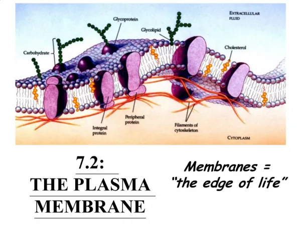
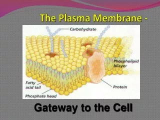
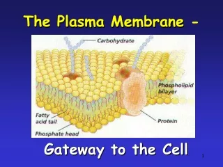
Why Cholesterol? • Importance in cell membrane integrity and signaling. • Regulates trans-membrane protein movement and plasma membrane-to-cytoskeleton attachment mechanics. Encyclopedia Britannica. 19 Aug 2009 <http://media-2.web.britannica.com/eb-media/74/53074-004-9F65D813.jpg>.
Objective We are testing the effect of various concentrations of cholesterol on plasma membrane biomechanics by pulling nanotubes (tethers) from the membrane and calculating the tether force.
Optical Tweezers • We are using optical tweezers to study plasma membrane biomechanics. • Our setup consists of a Solid-state diode pump laser (λ = 1064 nm), various optical components (100x objective), and a piezoelectric stage (nm resolution for both {x,y,z} movement and velocity).
Viscoelastic Model • The plasma membrane can be represented by a viscoelastic model. • The mechanics of the tethers are explained by a second-order Maxwellian spring – dash plot model of viscoelasticity. • The time-resolved effect on the tether force will be tested (more on this in the Methodology). Biophysical Journal 89(2005): 4090-4095.
Static Calibration Procedure • Trapping Force Calibration Materials: • DMEM Complete Media (Dulbecco’s Modified Eagle Medium with FBS and Penicillin/Strep.) Serum-enriched • Invitrogen™ Fluorescent Sulfate-Modified Beads (2 µm radius) • Piezoelectric stage • Power Meter • Calibration procedure: • Pass DMEM media through a trapped bead using a piezoelectric stage at various velocities (in µm/s) at various output power measurements (W) • Measure the velocity when the bead is dislodged from the trap. • Use Stokes’ Law to calculate the Escaping Force (pN) Fd= 6πηRV Where Fd = Viscous Drag Force, η = Viscosity, R = Radius of the Bead, and V = Escaping Velocity
Diagram Here, the diagram illustrates the bead becoming dislodged from the trap.
This calibration graph will be used to convert the output power into the tether force.
Cell Culturing Procedure • HEK 293 cells should be passaged when the flask/plate is ~80% confluenced. • DMEM Complete Medium and trypsin should be heated in a water bath for ~30 minutes before use to prevent thermal shock for the cells. • To passage: • Old media should be removed from the flask/plate • Cells should be washed with FBS to remove all of the old media, then FBS should be removed • 500 µL to 1 mL of trypsin should be added to the plate/flask, and should then be incubated for 1-2 minutes • Clusters of cells should be broken apart using both physical taps as well as rapid sucking in-and-out of 5 mL of new medium from the plate/flask. • Medium and trypsin in the plate/flask should be pipetted into another container, then distributed in different concentrations for different types of containers.
Cholesterol Manipulation Procedure • 3 mM and 5 mM concentrations. • Cholesterol depleted using M-β-CD (Methyl-Beta-Cyclodextrin) • Cholesterol enriched using water-soluble cholesterol obtained from Sigma-Aldrich™ in the form of cholesterol carrier – 51 mg cholesterol / 1 g of material. • The prepared media are vortexed for 3-4 minutes, followed by incubation for 30 minutes (37°C at 5% CO2) before experimentation.
Static Tether Force Measurement Procedure Step 1 Step 3 Step 4 Step 2
Very clear correlation: As cholesterol is depleted, the tether force increases, and as cholesterol is enriched, the tether force decreases. The means are also statistically significantly different.
Concentration-Dependent Conclusion • The tether forces increase as the cholesterol is depleted, and vice versa. • The order of tether forces is: Cholesterol-Depleted > Untreated > Cholesterol-Enriched • Statistically significant results • Also, the higher the concentration, the stronger the effect in either direction is – the elastic regime becomes more dominant as concentration increases.
Time-Resolved Results Conclusion • With no delay, the elastic regime more accurately portrays the peak tether forces. • As the viscous regime takes over, the tether forces are much lower, and are much less correlated. • This means that the viscous component of force is more dominant over time.
Future Research • Testing different tether pulling velocities. • Dynamic Force measurement. • Quantifying amount of cholesterol present.
Thanking: • N. Khatibzadeh • Dr. Sharad Gupta • O.S. Beane • Professor B. Anvari • Anvari Lab • J. Wang • National Science Foundation
Questions? Thanks for your time.