Download
1 / 29
360 likes | 772 Vues
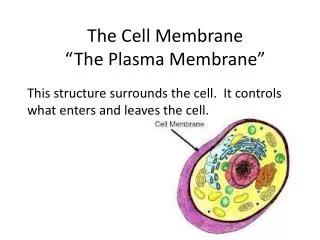
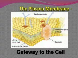
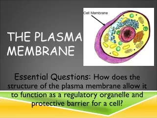
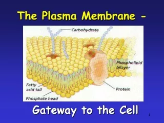
The Plasma Membrane. 13.1 Bilayer structure of the plasma membrane. Studies of the red blood cell plasma membrane provided the first evidence that biological membranes consist of lipid bilayers.

E N D
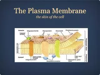
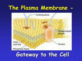
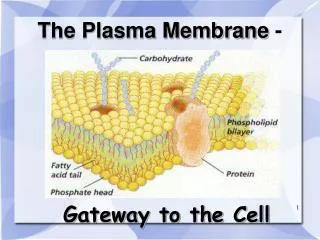
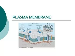
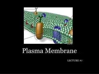
13.1 Bilayer structure of the plasma membrane • Studies of the red blood cell plasma membrane provided the first evidence that biological membranes consist of lipid bilayers. • The fundamental structure of the plasma membrane is the phospholipid bilayer, which forms a stable barrier between two aqueous compartments. • Proteins embedded within the phospholipid bilayer carry out the specific functions of the plasma membrane.
The Phospholipid Bilayer • Phosphatidylcholine -- glycerol phospholipid with a head group formed from choline. • Phosphatidylethanolamine -- glycerol phospholipid with a head group formed from ethanolamine. • Phosphatidylserine -- glycerol phospholipid with a head group formed from serine. • Sphingomyelin -- phospholipid consisting of two hydrocarbon chains bound to a polar head group containing serine.
The Phospholipid Bilayer • The fluid mosaic model of membrane structure is now generally accepted as the basic paradigm for the organization of all biological membranes. • Phosphatidylinositol is a phospholipid localized to the inner half of the plasma membrane, which plays an important role in cell signaling. • Glycolipids are lipids consisting of two hydrocarbon chains linked to a polar head group containing carbohydrates. • Cholesterol, a lipid consisting of four hydrocarbon rings, is a major membrane constituent of animal cells.
Membrane Proteins • Peripheral membrane proteins are proteins that dissociate from the membrane following treatments with polar reagents that do not disrupt the phospholipid bilayer. • Integral membrane proteins can be released only by treatments that disrupt the phospholipid bilayer. • Transmembrane proteins span the lipid bilayer with portions exposed on both sides of the membrane.
13.4 Solubilization of integral membrane proteins by detergents
13.6 Integral membrane proteins of red blood cells • The most abundant peripheral membrane protein of red blood cells is spectrin, which is the major cytoskeletal protein of erythrocytes. • There are two major integral membrane proteins in red blood cells—glycophorin and band 3.
Membrane Proteins • Some proteins are anchored in the plasma membrane by covalently attached lipids or glycolipids. • Glycosylphosphatidylinositol, or GPI, are glycolipids containing phosphatidylinositol that anchor proteins to the external face of the plasma membrane. • Other proteins are anchored to the inner leaflet of the plasma membrane by covalently attached lipids.
Mobility of Membrane Proteins • Lipid composition can perturb the free diffusion of membrane proteins. • The basolateral domain is the surface region of a polarized epithelial cell that is in contact with adjacent cells or the extracellular matrix. • The apical domain is the exposed free surface of a polarized epithelial cell.
13.13 The glycocalyx • The glycocalyx is formed by oligosaccharides of glycolipids and transmembrane glycoproteins. • Selectins are cell adhesion molecules that recognize oligosaccharides exposed on the cell surface.
13.15 Permeability of phospholipid bilayers • The internal composition of the cell is maintained because the plasma membrane is selectively permeable to small molecules. • Only small, relatively hydrophobic molecules are able to diffuse across a phospholipid bilayer at significant rates by using passive diffusion. • Passive diffusion is the simplest mechanism by which molecules can cross the plasma membrane. • Specific transport proteins mediate the selective passage of small molecules across the membrane, allowing the cell to control the composition of its cytoplasm.
Facilitated Diffusion and Carrier Proteins • Facilitated diffusion involves the movement of molecules in the direction determined by their relative concentrations inside and outside of the cell. • Carrier proteins bind specific molecules to be transported on one side of the membrane. • Channel proteins form open pores through the membrane, allowing the free diffusion of any molecule of the appropriate size and charge.
13.17 Model for the facilitated diffusion of glucose • Most cells, including erythrocytes, are exposed to extracellular glucose concentrations that are higher than those inside the cell, so facilitated diffusion results in the net inward transport of glucose.
13.18 Model of an ion channel • Porins permit the free passage of ions and small polar molecules through the outer membranes of bacteria. • Ion channels mediate the passage of ions across plasma membranes. • Ligand-gated channels open in response to the binding of neurotransmitters or other signaling molecules. • Voltage-gated channels open in response to changes in electric potential across the plasma membrane.
Ion Channels • Because ions are electrically charged, their transport results in the establishment of an electric gradient across the plasma membrane. • The Nernst equation is the relationship between ion concentration and membrane potential. • As action potentials, or nerve impulses, travel along axons, the membrane depolarizes.
Active Transport Driven by ATP Hydrolysis • Active transport, a process in which energy is provided by another coupled reaction, is used to drive the uphill transport of molecules in the energetically unfavorable direction. • Ion pumps responsible for maintaining gradients of ions across the plasma membrane, provide important examples of active transport driven directly by ATP hydrolysis.
Active Transport Driven by ATP Hydrolysis • ABC transporters belong to the largest family of membrane transporters and are characterized by highly conserved ATP-binding domains.
Active Transport Driven by Ion Gradients • Some molecules are transported against their concentration gradients using energy derived not from ATP hydrolysis, but from the coupled transport of a second molecule in the energetically favorable direction. • The epithelial cells lining the intestine provide a good example of active transport drive by the Na+ gradient.
Active Transport Driven by Ion Gradients • The flow of Na+ down its electrochemical gradient provides the energy required to take up dietary glucose and to accumulate high intracellular glucose concentrations. • A symport can transport two molecules in the same direction using the coordinated uptake of glucose and Na+.
Active Transport Driven by Ion Gradients • A uniport can transport only a single molecule using the facilitated diffusion of glucose. • An antiport uses active transport to move two molecules in opposite directions.
Endocytosis • Endocytosis is a process in which material to be internalized is surrounded by an area of the plasma membrane, which then buds off inside the cell to form a vesicle containing the invested material. • Phagocytosis, or cell eating, is the ingestion of large particles such as bacteria. • Pinocytosis, or cell drinking, is the uptake of fluids or macromolecules in small vesicles.
Phagocytosis • Phagolysosomes, which are phagosomes fused to lysosomes, contain lysosomal acid hydrolases that digest the ingested material. • The ingestion of large particles by phagocytosis plays distinct roles in different kinds of cells.
Receptor-Mediated Endocytosis • Receptor-mediated endocytosis, a form of pinocytosis, provides a mechanism for the selective uptake of specific macromolecules. • Clathrin-coated pits are specialized regions of the plasma membrane where specific cell surface receptors are found. • Dynamin, a membrane-associated GTP-binding protein, assists in the budding off of pits from the plasma membrane.
Receptor-Mediated Endocytosis • Clathrin assembles into a basketlike structure that distorts the membrane, forming invaginated pits. • Clathrin coated pits occupy about 1-2% of the surface area of plasma membranes.
Receptor-Mediated Endocytosis • Caveolae are small invaginations of the plasma membrane. • Caveolin is a protein that interacts with lipid rafts and forms caveolae. • Macropinocytosis is a process where large vesicles can mediate the uptake of fluids.
Protein Trafficking in Endocytosis • Endosomes are vesicles with tubular extensions, located at the periphery of the cell, that fuse with clathrin-coated vesicles which have shed their coats. • An important feature of early endosomes is that they maintain an acidic internal pH as the result of the action of a membrane H+ pump.
Protein Trafficking in Endocytosis • Recycling to the plasma membrane is the major fate of membrane proteins taken up by receptor-mediated endocytosis. • Ligands and membrane proteins destined for degradation in lysosomes are transported from early endosomes to late endosomes, which are located near the nucleus. • Receptor down-regulation is a phenomenon where receptor-ligand complexes are removed from the plasma membrane, thereby terminating the response of the cell to growth factor stimulation.