Download
1 / 13
130 likes | 145 Vues
This lecture covers the physical background of medical tomographies, including MRI mathematics, pulse methods for determining T2 and T1, magnetic resonance spectroscopy, and flow in MRI. It also discusses the advantages and disadvantages of fMRI and movie reconstruction from human brain activity using fMRI.

E N D
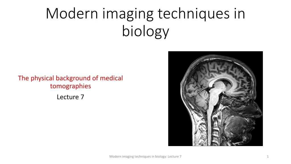
Modern imaging techniques in biology The physical background of medical tomographies Lecture 7 Modern imaging techniques in biology: Lecture 7
MRIthematics • Microscopic and macroscopic magnetization. The Bloch equation. T1 and T2 relaxation times. • Magnetic resonance. 90° pulse. FID: free induction decay. Effective T2. • Pulse method for determining T2 and T1. Spin echo. Magnetic resonance spectroscopy. • Imaging. Selective excitation and read out of a slice. • Pulse sequences and contrast. Flow in MRI. • fMRI. BOLD. Echo planar imaging (EPI). Spin echo (SE) and gradient echo (GE) for fast imaging. https://mri.byu.edu Modern imaging techniques in biology: Lecture 7
fMRIpros and cons Advantages: • non-invasive • fair spatial and temporal resolution • can image the whole brain almost simultaneously Disadvantages: • measuresindirectsignal (nottheelectricsignal of neuronsknownto be directlycorrelatedwithinformationprocessing, i.e., brainfunction) • measurestheintegralsignal of manyneurons Modern imaging techniques in biology: Lecture 7
Movie reconstruction from human brain activityusingfMRI Scientists use brain imaging to reveal the movies in our mind http://news.berkeley.edu/2011/09/22/brain-movies/ Modern imaging techniques in biology: Lecture 7
T1vs T2/T2* weigthedimages • Normalanatomicalimages of thebrainare most often T1-weighted. • fMRIimagesare T2or T2* weigthed. „A third commonly used sequence is the Fluid Attenuated Inversion Recovery (Flair). The Flair sequence is similar to a T2-weighted image except that the TE and TR times are very long. By doing so, abnormalities remain bright but normal CSF(cerebrospinal fluid) is attenuated and made dark. This sequence is very sensitive to pathology and makes the differentiation between CSF and an abnormality much easier.”http://casemed.case.edu Modern imaging techniques in biology: Lecture 7
fMRI: functional MRI Since1890 we knowthathemodynamics (blood flow, oxigenation) stronglycorrelateswithneuralactivity. 1-5 secs after an increasedneuronalactiviytheblood flow increases -> increasedoxigenation, increasedcerebralbloodvolume (CBV) Oxigentransport: O2 @redbloodcells (RBC) @Hemoglobin (Hb): Hemoglobin (Hb), Wikipedia Modern imaging techniques in biology: Lecture 7
MRI canmeasure • Tissueperfusion • Bloodoxygenation • Bloodvolume • Waterdiffusion FirstfMRIwasaquiredby a contrastagenttomeasure local CBV (cerebralbloodvolume). Then CBV withoutcontrastagent, and soonafterthatBOLDappeared. http://www.neurologyindia.com Modern imaging techniques in biology: Lecture 7
BOLD-contrast: Bloodoxigenationleveldependentcontrast Fe in theHemegroup is in high spin stateand thusparamagnetic in deoxy hemoglobin deoxy-Hb. 4 unpairedelectrons. http://mriquestions.com/bold-contrast.html BOLD since1990: hemodynamicresponse. Hemegroup: iron (Fe) ion held in a heterocyclic ring, known as a porphyrin Modern imaging techniques in biology: Lecture 7
Origin of BOLD contrast „The presence of paramagnetic deoxyhemoglobin within red blood cells creates local magnetic field distortions (susceptibility gradients) in and around blood vessels. These local field disturbances cause nearby stationary and slowly moving spins to have different resonance frequencies and phase shifts. The resultant intravoxel dephasing is a classic T2*-shortening effect most prominent near larger veins and accentuated by use of GRE sequences with echo times (TEs) close to T2*. The effect scales linearly with field strength (Bo) and is the dominant mechanism for BOLD contrast at 1.5T.” http://mriquestions.com/bold-contrast.html Paramagnetic deoxyhemoglobin (D) confined to red blood cells causes a local field distortion in and around the vessel. Modern imaging techniques in biology: Lecture 7
BOLD contrast:T2 and T2* Whereasmagneticsusceptibility of thediamagneticoxy-Hb is similartothesusceptibility of tissues,paramagneticdeoxy-Hb is different. The presence of deoxy-Hbresults in magneticfieldinhomogeneity. wherebz is the local fluctuatingmagneticfield. whereis thestaticfieldinhomogeneity. Both T2 and T2* willcorrelatestronglywiththe BOLD signal. Modern imaging techniques in biology: Lecture 7
Echo-planarimaging (EPI): MRI in a fraction of a second 1 excitation per image. A single RF shotcanscantheentirekspace. , Modern imaging techniques in biology: Lecture 7
Echo-planarimaging (EPI) Waystoscanthe k spaceor Fourier space. A singleRF pulsecanscantheentirekspace. M. K. Stehlinget al. Science 254, 43-50 (1991). Modern imaging techniques in biology: Lecture 7
Gradientecho (GE) versus spin echo (SE) The SE pulse sequence has a 90° excitation pulse (GE has a small excitation pulse), and SE refocuses some of the dephasing which occurs during the echo time using a 180° refocusing RF pulse. Because only one RF pulse is applied in GE, the echo can be recorded more quickly, resulting in a shorter echo time. If low flip angles are used, TR can also be shorter. ThusGE is preferred for rapid imaging techniques. GE image contrast is dictated by T2*, unlike in SE where image contrast is dictated by T2. In SE the signal-to-noise ratio is higher. http://www.revisemri.com Spin-echo (SE) versus gradientecho (GE) Modern imaging techniques in biology: Lecture 7