Download
1 / 75
750 likes | 778 Vues
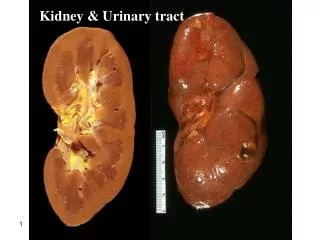
Investigation Of kidney and Urinary tract Dept. pathological physiology By Pavel Maruna. Fun ctional test s Glomerul ar filtra tion Tubul ar resorp tion C oncentr ation test Acidifi c a tion test Blood gases Imaging met h od s Native X-ray scintigra phy angiogra phy

E N D
Investigation Of kidney andUrinary tract Dept. pathological physiology By Pavel Maruna / 75
Functional tests Glomerular filtration Tubular resorption Concentration test Acidification test Bloodgases Imaging methods Native X-ray scintigraphy angiography Ultra-sonography MRI Endoscopy/ laparoscopy Laboratory tests / 75
Definitions Renal insuficiency Kidneys are able to maintain homeostasis of the inner milieu under normal conditions, but unable to do so in a stress conditions (infection, surgery, overlod by water and electrolytes). Renalfailure Kidneys are unable to maintain homeostasis of the inner milieu even under basic conditions. Uremia Syndrome of higher level of nitrogen metabolites in inner milieu, which can develop as a consequence of (mostly chronic) renal failure. / 75
Chronic renal failure/ uremic syndrome • 1. Retention of • water • electrolytes • small molecules • medium size molecules (500 - 3000 D) • 2. Losses of • water, electrolytes • amino acids, proteins, vitamines (soluble in water) • 3. Lower production of • Erythropoietin • 1,25-OH-D3 (vitamin D) • 4. Trade off hypothesis • = picture develops due to compensatory mechanisms / 75
Acute renalfailure According to site/ causes 1. Pre-renal 2. Renal (parenchymatous) 3. Post-renal (obstructive) / 75
Acute renal failure Causes 1. Prerenal Changes in haemodynamics Circulation shock, (Losses of blood, water and electrolytes) / 75
Acute renal failure Causes 2. Renal (parenchymatous) – mostly defects of tubules • TIN (tubulo-interstitial nephritis) • Glomerulo-nephritis (streptococcus, SLE, Goodpast. sy) • Nefrotoxic drugs (CCl4, etylenglykol, propylenglykol, Hg, Au,Bi, • nefrotox. substances (SFA, gentamycin, cefaloridin), amanita phal. • hemolysis (incompatible TRF) • crush syndrom • burns (dehydration, sepsis, toxemia) • toxo-infectious insult (sepsis) • childbirth, abortion, surgery • acute pancreatitis • Vascular disorders (occlusion a. renalis, • thrombosis of kidney veins, hypertension) / 75
Acute renal failure Obstruction of urinary tract Causes 3. Post-renal urolithiasis, blood clots tumours prostatic hypetrophy retro-peritoneal fibrosis surgical fixation of ureters atoniaof urinary bladder / 75
Acute renal failure Parameter Pre-renalf. Renal f. Holds only in a condition before cure (by diureticsand/or infusion). / 75
Acute renal failure Stages 1. Initial ... Dominated by its cause/ primary disease 2. Anuric / oliguric phase 3. Diuretic ... Polyuric phase (up to 5-6 l / 24 h) 4. Convalescence ... Sanatio ad integrum can take 1 year Oliguria < 500 ml / 24 h Anuria < 100 ml / 24 h / 75
Chronic renal insuficiency Final stage of various kidney ailments Staging I. Fullycompensated (cr. < 300) II. Compensated retention (cr. = 300-700) III. De-compensated retention (cr. > 700) ... Haemo-dialisys IV. Uremia 44 % glomerulonephritis, glomerulopathy 25 % TIN (tubulo-interstitial nephritis) 10 % kidney polycystosis / 75
Uremia • Water and electrolytedysbalance • losses / retention of water • Na (dilution, distribution, depletion) • K (retention) oedemas dehydration weakness, tiredness dyspepsia (anorexia, morning nausea, vomiting, diarrhea) arrythmia, perikarditis / 75
Uremia Bleeding Secondarygout Poly-neuropathy / 75
Uremia • Renal (reno-parenchymatous) • hypertension • Factors: • presoric (kidney hypoperfusion renin) • depresoric (kallikrein/kinins, PG E) • excretionof Na, H2O x Renovascular hypertension Renal presoric mechanisms (renin) only Hypo-perfusionin renal artery stenosis / 75
Uremia • Anemia • Factors: • Epo • vitamin losses, protein losses (proteinuria) • blood losses (hematuria) • low iron (inflammation, Trf) • toxic suppression of bone marrow • inflammatory inhibition of erythropoiesis / 75
Uremia • Renal osteodystrophy • Factors: • PO4 • 1,25-0H-vit. D3 ... Lower production • Ca ... losses, intestinal resorption • secondary, PTH ... bone resorption / 75
decreased production by the proximal convoluted tubule retention 1,25-0H-vit. D3 PO4 binding of serum Ca gut uptake of Ca renal loss Ca secondary hyperparatyreodidism PTH bone resorption, commonly from subperiosteal regions and tufts of the phalanges, proximal humerus, tibia and femur, and calvarium / 75
Uremia Renal osteodystrophy Higher para-thyroid activitycausing characteristic subperiostal resorption “Salt and pepper” skull / 75
Uremia Renal osteodystrophy Bone changes are partially reversible snapshots of the same finger before and 6 months after therapy of secondary hyper-parathyreosis / 75
Uremia Secondary infections Bronchitis, broncho-pneumonia Hepatitis Sepsis Cheyne - Stokes breathing pattern / 75
Acute dialysis Principle The dialysis membrane allows to exchange of low molecular substances to dialysis solution in accordance of its concentration (x molecules bound on plasma proteins) / 75
Acute dialysis Semipermeable dialysis membrane / 75
Acute dialysis The possible combination with ultrafiltration (in hyperhydratation, pulmonary edema) Utrafiltration The membrane process that uses moderate hydraulic pressure to transfer water and low molecular weight species through a membrane while retaining colloids and large organic molecules / 75
Acute dialysis Indications 1. Renal failure • uremia • anuria / oliguria > 3 days • creatinine > 700 umol / l • urea > 30 mmol / l • urea > 10 mmol / l / day • K > 6,5 mmol / l • acidosis • hyperhydration (conservatively immedicable) / 75
Acute dialysis Indications 1. Renal failure 2. Intoxication ... drugs non-bound on proteins psychiatric drugs fridex (coolant fluid) / 75
Acute dialysis Indications 1. Renal failure 2. Intoxication 3. Ca 4. urikemia ... e.g. after cytostatic therapy of leukemia 5. Hypotermia 6. Alcalosis ... rarely (not in CZ) / 75
Acute dialysis Contraindications: Only terminal stage of malignancy Not age or diagnosis All patients with creatinine > 300 nmol / L have to be followed in predialysis centers / 75
Peritoneal dialysis Principle Peritoneum is used as a dialysis membrane. The abdominal cavity is filled by a dialyze liquid. The liquid (artificial ascites) is get out after some interval. CAPD = Continual ambulatory peritoneal dialysis / 75
Hemoperfusion Principle The blood flows in hemoperfusion capsules through an absorbent. Elimination method of toxins bound on proteins hemoperfusion capsule / 75
Dialysis Complications Sy of insufficient dialysis Sy of excessive dialysis ... a loss of AA, vitamins, enzymes, hormones, hypotension due to hypovolemia Sy of disequilibrium ... a brain edema duo a quick dialysis Sy of hard water ... Ca in dialyse fluid hypertension, vomiting, fatigue, headache Infectious complication ... hepatitis B / 75
Urine biochemistry • U-Na ... 100 - 200 mmol / 24 h • U-K ... 30 - 80 mmol / 24 h • U-Na : U-K < 1 ... Na/K exchange in distal tubuli (aldosterone) • Pathology: primary kidney dis. • renin / angiotensin / aldosterone / 75
Urine biochemistry • Excretion fraction (EF) • = The fraction of its glomerular filtration flux, which passes to and is excreted in the urine • EF = Jexcr/Jfiltr • Jexcr = (Cu ×V°u) and Jfiltr =(GFR × Cfiltr). It follows that: • EF = (Cu ×V°u) /(GFR × Cfiltr) • Cfiltr = the concentration of the substance in the ultrafiltrate. • Inulin EF = 1. • Substances with an EF > 1 are subject to net secretion. • Substances with an EF < 1 are subject to net reabsorption. / 75
Urine biochemistry • Proteinuria • Physiol. < 150 mg / 24 h • Pathol. > 500 mg / 24 h • Haevy > 3500 mg / 24 h • Nephrotic sy > 5000 mg / 24 h • Methods of quantifying proteinuria: • urine dipstick test • sulfosalicylic acid tests Protein urinary excretion of > 2 g per 24 h is usually a result of glomerular disease. Young men with proteinuria < 2 g per 24 h and who have a normal creatinine clearance should be tested for orthostatic proteinuria / 75
Urine biochemistry • Proteinuria Glomerular x Tubular / 75
Urine biochemistry • Proteinuria Glomerular Increased glomerular capillary permeability to protein Primary or secondary glomerulopathy Tubular Decreased tubular reabsorption of proteins in glomerular filtrate Tubular or interstitial disease Overflow Increased production of low- molecular-weight proteins Monoclonal gammopathy, leukemia / 75
Urine biochemistry • Proteinuria • Nephrotic syndrome • Diagnostic criteria: • heavy proteinuria (>3500 g / 24 h) • hypoalbuminemia • edema • hyperlipidemia • lipiduria / 75
Urine biochemistry • Dipstick strip tests • pH • Glucose • Protein • Blood • Bilirubin • Urobilinogen • Ketones • Nitrite • Leukocytes / 75
Urine sediment • Addis: RBC < 2 mil. WBC < 4 mil. casts < 100 000 / 24 h • Hamburger: RBC < 2000 WBC < 4000 casts < 60 - 70 / min. • Phase-contrast RBC microscopy • ... to determine an origin of RBC • RBC from glomeruli ... deformation • RBC from urinary trct ... intact, smooth cells / 75
Urine sediment • Phase-contrast RBC microscopy Intact RBC (extraglomerular origin) Marginal deformation (RBC from glomeruli) / 75
Urine sediment RBC WBC Bacteria Squamous cells / 75
Urine sediment RBC casts WBC casts Granular casts Waxy casts / 75
Urine sediment Cystine crystals Tyrosine crystals / 75 Calcium oxalate crystals “Coffin lid” struvite crystals
Serum biochemistry • Urea • = A breakdown product of proteocatabolism • Serum urea depends upon both protein turnover and kidney function • Normal values: < 7,5 mmol / l • ... glomerular filtration rate (orient. parameter) • dehydration • proteocatabolism / 75
Serum biochemistry • Creatinine • = A breakdown product of creatine, which is an important part of muscle • Orientation parameter of GF • Normal values: • male < 124 umol / l • female < 115 umol / l (because of less muscle mass) / 75
Serum biochemistry • Creatinine • Creatinine x urea: • Stabile 24-h concentration • Independent on protein income • Independent on physical activity • Pathology: • ... glomerular filtration rate • muscular dystrophy, rhabdomyolysis • ... muscular dystrophy (late stage), myasthenia gravis / 75
Functional tests • Creatinine clearance • = GFR; Glomerular filtration rate • GFR = (Cu ×V°u) /Cp [(mg/ml)×(ml/min)/(mg/ml)= ml/min]. • = (U-creatinine x U-volume) / P-creatinine • = cca 2 ml / s (120 ml / min.) • Normal values: • male: 97 - 137 ml / min. • female: 88 - 128 ml / min. / 75
Functional tests • Creatinine clearance • Test compares the level of creatinine in urine with the creatinine level in the blood, usually based on measurements of • 24-h urine sample and • blood sample drawn at the end of the 24-h period / 75
Functional tests • Creatinine clearance • Pathology: • ... nephron damage • acute hemodynamic changes • ... oncotic pressure • glomerular membrane permeability (incip. DM nephropathy) / 75
Functional tests • Inulin clearance = The flux of inulin filtered through the glomerular barrier per min = GFR × Cp/0.94 Fructan molecule of the inulin / 75