Download
1 / 44
500 likes | 2.52k Vues
Extracorporeal Membrane Oxygenation (ECMO): Indications and Management Strategy. David Spielvogel, MD Surgical Director, Cardiac Transplant and Mechanical Circulatory Support Gilbert Tang, MD, MSc, MBA Cardiothoracic Surgeon, Transcatheter Heart Program

E N D
Extracorporeal Membrane Oxygenation (ECMO): Indications and Management Strategy David Spielvogel, MD Surgical Director, Cardiac Transplant and Mechanical Circulatory Support Gilbert Tang, MD, MSc, MBA Cardiothoracic Surgeon, Transcatheter Heart Program On behalf of the Cardiac Transplant and Mechanical Circulatory Support Team Westchester Medical Center, Valhalla, New York
OBJECTIVES • Understand the clinical indications for ECMO therapy • Identify procedural strategies and techniques of ECMO therapy • Discuss management strategy of ECMO in the ICU • Describe the ECMO experience at Westchester Medical Center
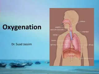
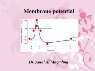
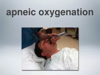
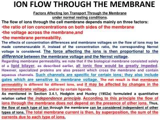
PHYSIOLOGY of ECMO Basic principle: De-saturated blood is drained via a venous cannula, CO2 is removed, O2 added through an “extracorporeal” device (an oxygenator), and the blood is then returned to systemic circulation via another vein (VV ECMO) or artery (VA ECMO)
VV ECMO • Perfusate blood returned to systemic circulation via venous cannula – travels into right ventricle and next pulmonary vasculature and is returned to the systemic circulation • Volume removed = volume returned; therefore no net effect on CVP, ventricular filling, or hemodynamics • CO2/O2 content in arterial blood supply is that of the blood arriving to right ventricle + any effects from gas exchange from remaining pulmonary function
VA ECMO • Replaces/augments both pulmonary and cardiac function • Perfusate mixes in the aorta with blood from left ventricle (arriving from compromised lungs); thus O2/CO2 content = content of blood returning from the circuit + that of pulmonary source; • Systemic blood flow = ECMO flow + pt’s own CO
Role of ECMO in Cardiogenic Shock • Bridge to recovery (BTR) • Bridge to decision (BTD) • Bridge to surgery • Bridge to long-term VAD • Bridge to transplant (BTT)
IABP in Cardiogenic Shock • Can initially stabilize patient • May not provide enough support • Requires a certain level of LV function • Limited by persistent tachycardia / arrhythmias • Does not unload the RV • Provides some pulsatile flow with ECMO
BRIDGE TO RECOVERY • Indications • Acute MI • Acute decompensated HF • Post-cardiotomy syndrome • Acute myocarditis • Severe rejection in transplant • Takotsubo’s • Massive PE • Respiratory failure and ARDS
BRIDGE TO SURGERY • Indications • Mechanical complications of AMI • VSD • Severe MR from papillary muscle rupture • CAD requiring CABG • Massive PE with heparin failure
BRIDGE TO Long-term VAD • Indications • Unable to wean off ECMO • Difficult donor match for transplant • Not a transplant candidate => LVAD as Destination Therapy
BRIDGE TO TRANSPLANT • Indications • Unable to wean off ECMO • Transplant candidate • Easy donor match for transplant
Predictors of Poor Outcomes • Multiorgan dysfunction • ARDS with sepsis • Severe neurological injury • Long time interval between shock and initiating ECMO
CONTRAINDICATIONS • Major CNS injury • Severe anoxia • Embolic or hemorrhagic stroke • Intracerebral hemorrhage • Multiorgan failure • Metastatic disease • Overwhelming sepsis
TWO TYPES OF ECMO: • Veno-arterial bypass - supports the heart and lungs • Veno-venous bypass – supports the lungs only
Equipment: Cannulas • VV ECMO: • Jugular vein, femoral vein • VA ECMO • Vein: femoral • Artery: • Femoral • Axillary • Aorta
Equipment: Pump, Oxygenator • ThoratecCentrimag pump & motor • Centrimag console • MaquetQuadrox oxygenator
PA Catheter CENTRIMAG IABP QUADROX OXYGENATOR Venous: percutaneous • Arterial: • Femoral percutaneous • Axillary graft • Aorta direct
R axillary artery R femoral vein
Axillary vs Femoral Cannulation AXILLARY FEMORAL • Side-arm graft sewn on • Antegrade perfusion better for cerebral and aortic root oxygenation, especially when lungs not oxygenating • Increased afterload • Risk of arm hyper-perfusion • Percutaneous • Need antegrade stick for forward perfusion • Retrograde perfusion increases atheroembolic risk • Ad-mixing with cardiopulmonary circulation => indequate cerebral and aortic root oxygenation if lungs not oxygenating
Check arterial line pressure! • High line pressure risks hyperperfusion and bleeding at axillary site • Need to Y the arterial outflow: • Bi-axillary • Axillary + femoral • Indications • Patients with large BSA • Small axillary artery
Anticoagulation • IV Heparin, target ACT of 200-240 seconds to prevent clotting upon interference of blood with prosthetic surfaces and in stagnant areas. • If high bleeding risk, ACT 180-220 s • Watch for platelet drop and heparin induced thrombocytopenia (HIT)
Monitoring an ECMO patient • Continuous cerebral SaO2 • CVP, PAP, CO • CXR – assess pulmonary edema • SvO2: 75% in VA ECMO and 85-90% on VV ECMO considered adequate as long as CO normal • EtCO2 – measures return of native lung function • aBG, lactate – tissue perfusion • Urine output, fluid balance – renal function • Labs: renal, hepatic function • Platelet count
POTENTIAL RISKS • Infection • Bleeding • Brain • Surgical site • Non-pulsatile flow • Renal insufficiency • Peripheral ischemia • Limb complications • Arm hyperperfusion • Leg ischemia • Air in circuit • Pump malfunction • Clots in the circuits • Heat exchanger malfunction • Cannula dislodgement
Criteria for Weaning ECMO • Pulmonary edema resolved • Minimal inotropes / pressors • End-organ dysfunction nearly recovered
ECMO Weaning Protocol • ICU • ECMO flow down to 1-1.5 L/min for 5 min • Assess CVP, PAP, CO • TTE to assess LV, RV function • OR • 3000-5000 U heparin • ECMO flow down to 1 L/min • Assess CVP, PAP, CO • TEE to assess LV, RV function, septal position • Explant ECMO if appropriate
Special Note on ECMO & LVAD • Pts with LVAD need to balance flow with both LVAD and ECMO to optimize end-organ perfusion • TEE to check septal position, need to unload RV • After ECMO explant, LVAD flow needs to increase b/c of LV preload increases
CONCLUSIONS • Rapidly evolving technology • Increasing array of indications • Excellent “tool” for ACS with cardiogenic shock • Shifting the paradigm of “bridge to recovery” • Presently investigating the “science” behind the clinical results