Download
1 / 9
100 likes | 483 Vues
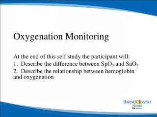
Oxygenation Monitoring. At the end of this self study the participant will: 1. Describe the difference between SpO 2 and SaO 2 2. Describe the relationship between hemoglobin and oxygenation. Pulse Oximetry (SpO 2 ). Adequate Tissue Oxygenation depends on three things:

E N D
Oxygenation Monitoring At the end of this self study the participant will: 1. Describe the difference between SpO2 and SaO2 2. Describe the relationship between hemoglobin and oxygenation
Pulse Oximetry (SpO2) • Adequate Tissue Oxygenation depends on three things: • Adequate Oxygenation – look at the PaO2 • Adequate Hemoglobin – to carry the O2 • Adequate Circulation • Pulse Oximetry measures: • The ability of the blood to bind to oxygen • Gas exchange in the lungs • Pulse Oximetry does not measure the oxygen delivery to the tissues
Hemoglobin’s role • Hemoglobin • oxygen carrier • normal 12-16 gm/dl • Critical threshold = less than 10 gm/dl • Physician alert value <4 gm/dl • High binding capacity with • oxygen • carbon dioxide • carbon monoxide
Oxyhemoglobin = functional oxygen • % of available oxygen sites on hemoglobin that are attached to an oxygen molecule • Oxygen Saturation • normal 95-100% • critical threshold normally <90% • SaO2 is Arterial saturation • SpO2 is Pulse oximetry • Skin color is NOT a reliable indicator of oxygenation Central Cyanosis
Dysfunctional Hemoglobin • 97% of oxygen is attached to hemoglobin • 2-4% is normally dysfunctional • Carboxyhemoglobin (COHb) • Carbon monoxide has a greater infinity to bind to Hgb than oxygen, therefore the oxygen carrying capacity of Hgb is reduced • Expected values: • 0.5-1.5% (non-smokers)1.0-5.0% (average smokers)5.0-9.0% (heavy smokers) • Physician alert value (automatic callback): ≥20%
Dysfunctional Hemoglobin • Methemoglobin (MetHb) • heme protein changes state (ferrous to ferric)- thus is incapable of binding with oxygen • Normal <2% • Physician alert value (automatic call-back): ≥5.0% • Causes include • Local anesthetics • Environmental 10-20% metHb - Skin discoloration only (most notably on mucus membranes) 20-30% metHb - Anxiety, headache, dyspnea on exertion 30-50% metHb - Fatigue, confusion, dizziness, tachypnea, palpitations 50-70% metHb - Coma, seizures, arrhythmias, acidosis Greater than 70% metHb - Death
Pulse Oximetry (SpO2) • Process • Light shone through tissue • Oxygenated and reduced hemoglobin absorbs different amount of light • amount of light detected on other side of tissue determines SpO2 • Indirect measurement • Is an estimation of SaO2
SpO2 • Normal 95-100% • Critical Threshold <90% • Should correlate with SaO2 • If correlation present, can use for ventilator effectiveness
Pulse Oximetry LimitationsMay cause inaccurate readings • Compression of artery • during BP • External Light Sources • IV dyes • Nail polish • Inconsistent pulse detection • Hyperlipidemia or hyperbilirubinemia • Low perfusion state • shock • Vasoconstriction • vasopressors, hypothermia • Dysfunctional hemoglobin • carboxyhemoglobin, methemoglobin • Motion artifact