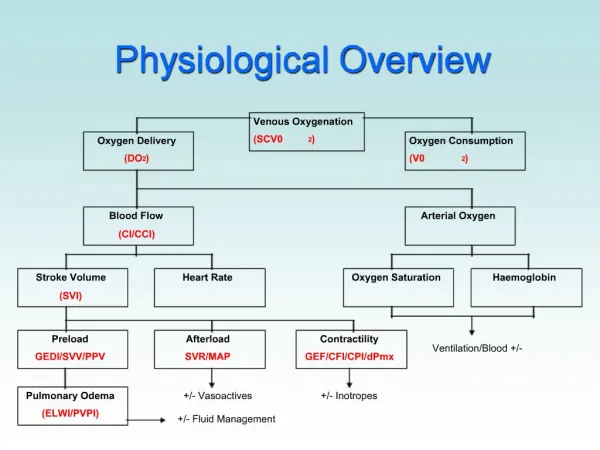
Improving Oxygenation
Improving Oxygenation. Chapter 14. Oxygenation. Assessed by FiO2, SaO2, PaO2, Hb Ideal to keep FiO2 < .4/.5, PaO2 60-90 mmHg, and CaO2 20mL/dL The SpO2 can be used to titrate FiO2; goal is >90% FiO2 may be adjusted using the following equation: Desired FiO2 = PaO2 desired X FiO2 known

Improving Oxygenation
E N D
Presentation Transcript
Improving Oxygenation Chapter 14
Oxygenation • Assessed by FiO2, SaO2, PaO2, Hb • Ideal to keep FiO2 < .4/.5, PaO2 60-90 mmHg, and CaO2 20mL/dL • The SpO2 can be used to titrate FiO2; goal is >90% • FiO2 may be adjusted using the following equation: Desired FiO2 = PaO2 desired X FiO2 known PaO2 known
A patient with myasthenia gravis is started on mechanical ventilation. The CXR is normal. Breath sounds are clear. Initial ABG’s on .25 FiO2 after 20 minutes on the ventilator are 7.31/62/58/31. What changes in ventilator settings might improve this patient's ABG findings? This patient has respiratory acidosis. The PaO2 indicates moderate hypoxemia. A common reaction by clinicians in this situation is to increase the FiO2. However the cause of the hypoxemia is the elevated PaCO2. An increase in CO2 of 1mmHg reduces the O2 by 1.25mmHg. The PaCO2 is about 40mmHg above normal therefore the PaO2 will be about 50mmHg below its actual value. The most appropriate action is to increase ventilation Clinical Rounds 14-1, p. 296
Selection of FiO2 • Levels >0.6 can result in oxygen toxicity • 100% Oxygen can cause the rapid formation of absorption atelectasis and increase pulmonary shunting • When PaO2 remains low on high FiO2 significant shunting, V/Q abnormalities and/or diffusion defects are present
After being supported on a ventilator for 30 minutes, a patient's PaO2 is 40mmHg on an FiO2 of 0.75. Acid-base status is normal and all other ventilator parameters are within the acceptable range. PEEP is 3 cmH2O. What FiO2 is required to achieve a desired PaO2 of 60 mmHg? Is this possible? Can you think of another form of therapy to improve oxygenation? Desired FiO2 = (60x0.75)/40= 1.13 You cannot give more than 100% O2. The appropriate change is the FiO2 to 100% and increasing PEEP Clinical Rounds 14-2, p. 298
Strategies to Improve Oxygenation • Increase the mean airway pressure • PIP • Total PEEP • I:E ratios • Respiratory rate • Inspiratory flow pattern • Paw affects mean alveolar pressure and alveolar recruitment and therefore oxygenation Figure 14-01. A pressure-time waveform illustrating mean airway pressure (aw). Vertical lines under the pressure-time curve represent frequent readings of pressure over the total respiratory cycle. The sum of these pressure readings (i.e., the area under the curve) divided by the cycle time will give the value for mean airway pressure. (See text for additional information.)
Goals of PEEP • Enhance tissue oxygenation • Maintain a PaO2 > 60mmHg and SpO2 >90% at an acceptable pH • Recruit alveoli and maintain them in an aerated state • Restore FRC • Opportunity to decrease FiO2 to safer levels
Atelectasis • Partial or complete collapse of alveoli • Result of: • Blocked airways • Shallow breathing • Sufactant deficiency • Treat what is causing the problem
Interface • Mask CPAP • Nasal CPAP • Endotracheal or Tracheostomy tubes • Flow resistors • Threshold resistors Free standing CPAP systems
PEEP Ranges • Minimum or Low PEEP • 3-5cmH2O • Preserves normal FRC • Therapeutic PEEP • >/= 5cmH2O • Used to treat refractory hypoxemia • High levels are only beneficial to a small % • Associated with cardiopulmonary complications • Optimum of Best PEEP • Level at which the maximum beneficial effects of PEEP occur and is not associated with profound cardiopulmonary side effects and it is accomplished at safe FiO2 levels
Indications for PEEP/CPAP • Bilateral infiltrates on CXR • Recurrent atelectasis with low FRC • Reduced lung compliance • PaO2 <60mmHg on high FiO2 >0.5 • PaO2/FiO2 ratio <200 for ARDS and <300 for ALI • Refractory hypoxemia: PaO2 increases 10mmHg with FiO2 increase of 0.2
Specific Disorders that benefit from PEEP • ALI • ARDS • Cardiogenic pulmonary edema • Bilateral diffuse pneumonia
Initiating PEEP • PEEP should be started as soon as possible • Best to look at several factors when deciding if the best PEEP level has been achieved • Increases in PEEP are generally done in 3-5cmH2O in adults 2-3cmH2O in infants • Cardiovascular status is closely monitored
Optimum PEEP study • Reserved for patients requiring a PEEP of 10cmH2O or greater • Extensive monitoring during the study • Target Goals: • A PaO2 of 60mmHg on FiO2 <0.4 • Optimum oxygen transport is present • A shunt of less than 15% • A minimal amount of cardiovascular compromise – adequate BP, decrease of <20% cardiac output and stable pulmonary vascular pressures • Improving lung compliance and improved lung aeration • A PaO2/FiO2 ratio of more than 300 • The point of minimum arterial to end-tidal PCO2 gradient • Optimum mixed venous oxygen values
Figure 14-2 A, The stiff lungs and increased shunt result in a drop in FRC and PaO2. B and C, as PEEP is increased, CS and PaO2 improve as the FRC increases, resulting in a lowering of the shunt effect. D, Too much PEEP has been used, and CS and cardiac output decrease as the FRC is increased above the optimum level.
Assessment during PEEP Study • Patient Appearance • Blood Pressure • Breath Sounds • Ventilator Parameters • Static Compliance • PaO2/FiO2 • Adequacy of ventilation • P(A-a)O2 • P(a-et)CO2 • Hemodynamics • C(a-v)O2 • PvO2 • Cardiac Output
Contraindications for PEEP • Hypovolemia • Untreated or significant pneumothorax • Elevated ICP • Pre-existing hyperinflation – emphysema • Unilateral lung disorders Overdistention vs hyperinflation
Weaning from PEEP • Exact length of time PEEP is required is not known • Trial reductions can be attempted when: • Patient demonstrates an acceptable PaO2 on an FiO2 <0.40 • Patient is hemodynamically stable and nonseptic • Patient's lung condition should have improved
Recruitment Maneuvers • A sustained increase in pressure in the lungs with the goal of opening as many collapsed lung units as possible • Once recruited the lungs are kept open by maintaining an adequate PEEP • Consists of three parts • An inflation maneuver to open as much of the lung as possible • A deflation maneuver to determine the point at which a majority of the lung begins to collapse • Another inflation recruitment maneuver to reopen the lung following its collapse
Hazards of Recruitment Maneuvers • Significant increases in thoracic pressure for an extended period of time can result in: • Decreased venous return • Drop in cardiac output • Drop in BP • Uneven effects in the lungs • Variability among patients
Recruitment Maneuvers Types of RM Summary ↑ oxygenation, ↓ shunt, ↑ pulmonary compliance Work early in ARDS No uniform way of performing this maneuver May reduce atelectasis post-op Generally safe Important to set PEEP to prevent alveolar collapse post RM • Sustained Inflation • PCV with high PEEP • PCV with increased PEEP • Sighs