Oxygenation
Oxygenation Unit Eight Respiratory system Oxygen: a clear, odorless gas that constitutes approximately 21 percent of the air we breathe for necessary all living cell. Respiration: is the process of gas exchange between individual and the environment.

Oxygenation
E N D
Presentation Transcript
Oxygenation Unit Eight
Oxygen: a clear, odorless gas that constitutes approximately 21 percent of the air we breathe for necessary all living cell. • Respiration: is the process of gas exchange between individual and the environment.
The process of respiration involves several components: • Pulmonary ventilation: the movement of air between the atmosphere and alveoli of the lungs. • Diffusion of oxygen and carbon dioxide between alveoli and capillaries. • Transport of oxygen and carbon dioxide via blood to tissues. • Diffusion of oxygen and carbon dioxide between capillaries and cell.
Functions of pulmonary system: • Ventilation: is the movement of air in and out of the lung. • Respiration: is the process of gas exchange.
Manifestations of Altered Respiratory Function • Cough • Sputum Production • Shortness of Breath • Chest Pain • Abnormal Breath Sounds • Accessory Muscle Use • Cyanosis • Clubbing fingers.
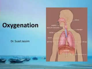
Anatomy and physiology of respiratory system: • 1) Upper respiratory tract: • a) Nose – made of cartilage and bone and is designed to warm, moisten, and filter air as it comes into the system. • b) Pharynx – (throat) conducts food and air.
Cont • C) Larynx – (voice box) connects the pharynx and the trachea; made of cartilage; contains vocal cords. • D) Epiglottis – flap of tissue that covers trachea; ensures food travels down the esophagus.
Alveolar sac Alveoli
Cont • 2) Lower Respiratory tract: • Trachea – (windpipe) tubular passage way for air; carries air to the lungs, C-shaped cartilage rings, divides at end. • Bronchi – pair of tubes that branch from trachea and enter lungs; have cartilage, lining is ciliated & secretes mucus. • Bronchioles – tiny tubes lacking cartilage and cilia; possess smooth muscle
Alveoli – cup shaped structures at the end of the bronchioles that resemble bunches of grapes; are in direct contact with capillaries (gas exchange); covered with SURFACTANT that prevent alveoli from collapsing. • Lungs – paired, cone-shaped organs that are surrounded by a pleural membrane, made of elastic tissue, and divided into lobes
Mechanics of Breathing • Inhaling (active process) – Air moves in. Why? • Gases move from an area of high pressure to low pressure • During inspiration – diaphragm pulls down and lungs expand • When lungs expand, it increase the volume, which decrease the pressure inside lungs
Lung pressure is lower than outside pressure, so air moves in. • Exhaling (passive process) – breathing out • Diaphragm and muscles relax • Volume in lungs and chest cavity decreases, so now pressure inside increases. • Air moves out because pressure inside is HIGHER than OUTSIDE atmosphere.
Respiration: • Exchange of O2 and CO2 between alveoli and blood • Partial pressure of O2 higher in alveoli than blood so O2 diffuses into blood • Partial pressure of CO2 higher in blood than alveoli, so CO2 moves into alveoli in opposite direction and gets exhaled out
Internal respiration • Internal respiration is exchange of O2 and CO2 between blood and tissues • Pressure of O2 higher in blood than tissues so O2 gets release into tissues. • Pressure of CO2 higher in tissue than in blood so CO2 diffused in opposite direction into blood. • CO2 Is a waste product. • O2 Is used in cellular respiration
3 Muscle Groups of Inhalation • Diaphragm: • contraction draws air into lungs • 75% of normal air movement • External intercostals muscles: • assist inhalation • 25% of normal air movement • Accessory muscles assist in elevating ribs: • sternocleidomastiod • serratus anterior • pectoralis minor • scalene muscles
Control of Breathing • Breathing is regulated by the rhythmicity center in the medulla and pons in brain stem. • Carotid body is sensitive to level of oxygen. • medulla rate and depth of breathing
Factor effecting oxygenation: • Environment: high altitude increase respiratory rate. • Exercise: physical exercise lead to increase respiratory rate. • Life style: smoking, occupation. • Health status: disease of cardiovascular disease. • Narcotics: morphine decrease respiratory rate. • Stress and anxiety.
Respiratory alteration: • Hypoxia: is condition of insufficient oxygen anywhere in the body from the inspired gas to the tissue. Cerebral function can tolerate hypoxia for only 3 to 5 min before permanent damage.
Sign of hypoxia: • Rapid pulse. • Rapid shallow respiration. • Increase restlessness. • Flaring nares. • Cyanosis.
Hypoventilation: inadequate alveolar ventilation can lead to hypoxia may result from disease of respiratory muscle, drug, and anesthesia. • Hypercabnia: accumulation of carbon dioxide in the blood. • Cyanosis: bluish discoloration of the skin nails beds and mucosal membrane
Altered breathing pattern: • Breathing pattern: rate, volume, rhythm, effort of respiration. • Normal respiration: (Eupnea) quite, rhythmic and effortless. • Tachypnea: rapid rate is seen with fevers, metabolic acidosis, pain and Hypercabnia. • Bradypnea: slow respiration rate, seen with narcotics and increase intracranial pressure from brain injury.
Hyperventilation: increase movement of air into and out of the lung. • Dyspnea: difficult of breathing. • Orthopnea: in ability to breathe except in an upright position
Obstructed air way: • Partially or completely in upper and lower respiratory tract.
Assessment • Nursing history: • Respiratory problem, cardiac problem, life style, cough and sputum. • Physical assessment: • Inspection, palpation, percussion and auscultation.
Diagnostic studies: • Sputum specimen, throat culture, arterial blood gases. • X- Ray examination. • Bronchoscopy and laryngoscopy. • Pulse oximetry: non invasive device measuring oxygen saturation.
Sputum collected for the following reason: • Culture and sensitivity: for identify a specific microorganism. • Cytology: to identify the origin, structure, function and pathology cell. • Acid bacillus: to identify the presence of tuberculosis.
Nursing diagnosis: • Ineffective air way clearance related to accumulation of secretion. • Ineffective breathing pattern related to dyspnea. • Altered tissue perfusion related to decrease cardiac out put. • Anxiety related to ineffective air way clearance.
Implementation: • Positioning the client to allow to maximum chest expansion. • Encourage frequent changes in position. • Encourage ambulating. • Deep breathing exercise and coughing. • Hydration to maintain moisturing of respiratory tract mucous membrane and easily to move respiratory secretion and decease incidence of infection.
Oxygen Administration • 1: Nasal Cannula. • 2: Face Mask. • 3: Oxygen Tent (for children). • 4: Venturi Mask. • 5: Non rebreather Mask. • 6: Partial Rebreather Mask. • 7: Endotracheal Tube (ETT).
The End Good Luck