Download
1 / 59
590 likes | 604 Vues
Dive into the world of cells with this guide covering microscopes, organelles, and the difference between prokaryotic and eukaryotic cells. Understand the structure and functions of the nucleus, endoplasmic reticulum, and more!

E N D
The Cell Organelles: Acceleration in 7th Grade Life Science
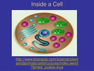
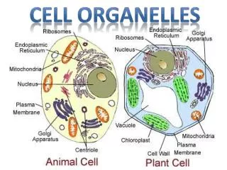
Cell: • A basic unit of living matter separated from its environment by a plasma membrane. • The smallest structural unit of life.
The Cell Theory • All living things ( organisms ) are made of one or more cells. • The cell is the basic unit of life ( it is the basic structure and carries out the basic functions of all organisms). • All new cells come from preexisting cells.
Microscope Features • Magnification: • Increase in apparent size of an object. • Ratio of image size to specimen size.
Microscope Features • Resolving power: • Measures clarity of image. • Ability to see fine detail. • Ability to distinguish two objects as separate. • Minimum distance between 2 points at which they can be distinguished as separate and distinct.
Microscopes • Light Microscopes: • Earliest microscopes used. • Lenses pass visible light through a specimen. • Magnification: Highest possible from 1000 X to 2000 X. • Resolving power: Up to 0.2 mm (1 mm = 1/1000 mm).
Types of Microscope • Electron Microscopes: • Developed in 1950s. • Electron beam passes through specimen. • Magnification: Up to 200,000 X. • Resolving power: Up to 0.2 nm (1nm = 1/1,000,000 mm).
Types of Microscope • Electron Microscopes: • Two types of electron microscopes: • 1. Scanning Electron Microscope: Used to study cell or virus surfaces. • 2. Transmission Electron Microscope: Used to study internal cell structures.
Components of All Cells: • 1. Plasma membrane: Separates cell contents from outside environment. Made up of phospholipid bilayers and proteins. • 2. Cytoplasm: Liquid, jelly-like material inside cell. • 3. Ribosomes: Necessary for protein synthesis.
Prokaryotic versus Eukaryotic Cells: • Feature Prokaryotic Eukaryotic • Organisms Bacteria All others (animals, plants, • fungi, and protozoa) • Nucleus Absent Present • DNA One chromosome Multiple chromosomes • Size Small (1-10 um) Large (10 or more um) • Membrane Absent Present (mitochondria, • Bound golgi, chloroplasts, etc.) • Organelles • Division Rapid process Complex process • (Binary fission) (Mitosis and Cytokinesis – Cell Cycle)
Relative Sizes of Prokaryotic and Eukaryotic Cells and Viruses
Relative Sizes of Structures • 1 nanometer (10-9 m) water molecule • 10 nanometers (10-8 m) small protein • 100 nanometers (10-7 m) HIV virus • 1 micron (10-6 m) cell vacuole • 10 microns (10-5 m) bacterium • 100 microns (10-4 m) large plant cell • 1 millimeter (10-3 m) single cell embryo • http://learn.genetics.utah.edu/content/begin/cells/scale/
Prokaryotic Cells • Bacteria and blue-green algae. • Small size: Range from 1- 10 micrometers in length. About one tenth of eukaryotic cell. • No nucleus: DNA in cytoplasm or nucleoid region. • Ribosomes are used to make proteins. • Cell wall: Hard shell around membrane. • Other structures that may be present: • Capsule: Protective, outer sticky layer. May be used for attachment or to evade immune system. • Pili: Hair-like projections (attachment). • Flagellum: Longer whip-like projection (movement).
Prokaryotic Cells: Lack a Nucleus and other Membrane Bound Organelles
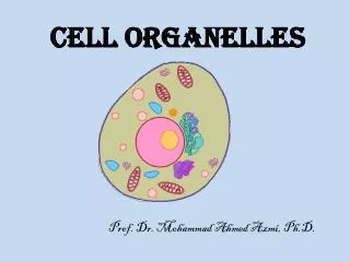
Eukaryotic Cells • Include protist, fungi, plant, and animal cells. • Nucleus: Protects and houses DNA. • Membrane-bound Organelles: Internal structures with specific functions. • Separate and store compounds. • Store energy. • Work surfaces. • Maintain concentration gradients.
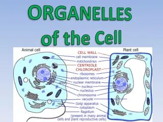
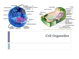
Membrane-Bound Organelles of Eukaryotic Cells • Nucleus • Rough Endoplasmic Reticulum (RER) • Smooth Endoplasmic Reticulum (SER) • Golgi Apparatus • Lysosomes • Vacuoles • Chloroplasts • Mitochondria
Nucleus • Structure: • Double nuclear membrane (envelope). • Large nuclear pores. • DNA (genetic material) is combined with histones and exists in two forms: • Chromatin (Loose, threadlike DNA, most of cell life). • Chromosomes (Tightly packaged DNA. Found only during cell division). • Nucleolus: Dense region where ribosomes are made.
Nucleus • Functions: • House and protect cell’s genetic information (DNA) • Ribosome synthesis
Endoplasmic Reticulum (ER) • “Network within the cell.” • Extensive maze of membranes that branches throughout cytoplasm. • ER is continuous with plasma membrane and outer nucleus membrane. • Two types of ER: • Rough Endoplasmic Reticulum (RER) • Smooth Endoplasmic Reticulum (SER)
Rough Endoplasmic Reticulum (RER) • Flat, interconnected, rough membrane sacs. • “Rough”: Outer walls are covered with ribosomes. • Ribosomes: Protein making “machines.” • May exist free in cytoplasm or attached to ER. • RER Functions: • Synthesis of cell and organelle membranes. • Synthesis and modification of proteins. • Packaging, and transport of proteins that are secreted from the cell. • Example: Antibodies
Smooth Endoplasmic Reticulum (SER) • Network of interconnected tubular smooth membranes. • “Smooth”: No ribosomes. • SER Functions: • Synthesis of phospholipids, fatty acids, and steroids (sex hormones). • Breakdown of toxic compounds (drugs, alcohol, amphetamines, sedatives, antibiotics, etc.). • Helps develop tolerance to drugs and alcohol. • Regulates levels of sugar released from liver into the blood. • Calcium storage for cell and muscle contraction.
Golgi Apparatus • Stacks of flattened membrane sacs that may be distended in certain regions. Sacs are not interconnected. • First described in 1898 by Camillo Golgi (Italy). • Works closely with the ER to secrete proteins. • Golgi Functions: • Receiving side receives proteins in transport vesicles from ER. • Modifies proteins into final shape, sorts, and labels proteins for proper transport. • Shipping side packages and sends proteins to cell membrane for export or to other parts of the cell. • Packages digestive enzymes in lysosomes.
The Golgi Apparatus: Receiving, Processing, and Shipping of Proteins
Lysosomes • Small vesicles released from Golgi containing at least 40 different digestive enzymes, which can break down carbohydrates, proteins, lipids, and nucleic acids. • Optimal pH for enzymes is about 5. • Found mainly in animal cells. • Lysosome Functions: • Molecular garbage dump and recycler of macromolecules (e.g.: proteins). • Destruction of foreign material, bacteria, viruses, and old or damaged cell components. • Digestion of food particles taken in by cell. • After cell dies, lysosomal membrane breaks down, causing rapid self-destruction.
Lysosomes, Aging, and Disease • As we get older, our lysosomes become leaky, releasing enzymes which cause tissue damage and inflammation. • Example: Cartilage damage in arthritis. • Steroids or cortisone-like anti-inflammatory agents stabilize lysosomal membranes, but have other undesirable effects (affect immune function). • Diseases from “mutant” lysosome enzymes are usually fatal: • Pompe’s disease: Defective glycogen breakdown in liver. • Tay-Sachs disease: Defective lipid breakdown in brain. Common genetic disorder among Jewish people.
Vacuoles • Membrane bound sac. • Different sizes, shapes, and functions: • Central vacuole: In plant cells. Store starch, water, pigments, poisons, and wastes. May occupy up to 90% of cell volume. • Contractile vacuole: Regulate water balance, by removing excess water from cell. Found in many aquatic protists. • Food or Digestion Vacuole: Engulf nutrients in many protozoa (protists). Fuse with lysosomes to digest food particles.
Interactions Between Membrane Bound Organelles of Eukaryotic Cells
Chloroplasts • Site of photosynthesis in plants and algae. • CO2 + H2O + Sun Light -----> Sugar + O2 • Number may range from 1 to over 100 per cell. • Disc shaped structure with three different membrane systems: • 1. Outer membrane: Covers chloroplast surface. • 2. Inner membrane: Contains enzymes needed to make glucose during photosynthesis. Encloses stroma (liquid) and thylakoid membranes. • 3. Thylakoid membranes: Contain chlorophyll, green pigment that traps solar energy. Organized in stacks called grana.
Chloroplasts Trap Solar Energy and Convert it to Chemical Energy
Chloroplasts • Contain their own DNA, ribosomes, and make some proteins. • Can divide to form daughter chloroplasts. • Plastid: Organelle that produces and stores food in plant and algae cells. • Other plastids include: • Leukoplasts: Store starch. • Chromoplasts: Store other pigments that give plants and flowers color.
Mitochondria (Sing. Mitochondrion) • Site of cellular respiration: • Food (sugar) + O2 -----> CO2 + H2O + ATP • Change chemical energy of molecules into the useable energy of the ATP molecule. • Oval or sausage shaped. • Contain their own DNA, ribosomes, and make some proteins. • Can divide to form daughter mitochondria. • Structure: • Inner and outer membranes. • Intermembrane space • Cristae (inner membrane extensions) • Matrix (inner liquid)
Origin of Eukaryotic Cells • Endosymbiont Theory: Belief that chloroplasts and mitochondria were at one point independentcells that entered and remained inside a larger cell. • Both organelles contain their own DNA • Have their own ribosomes and make their own proteins. • Replicate independently from cell, by binary fission. • Symbiotic relationship: the larger cell obtains energy or nutrients and the smaller cell is protected by larger cell.
Cytoplasm DNA Plasma membrane Ancestral prokaryote Infolding of plasma membrane Nucleus Endoplasmic reticulum Nuclear envelope Engulfing of aerobic heterotrophic prokaryote Cell with nucleus and endomembrane system Mitochondrion Mitochondrion Engulfing of photosynthetic prokaryote in some cells Ancestral heterotrophic eukaryote Plastid Ancestral Photosynthetic eukaryote Figure 26.13 Endosymbiont Theory
The Cytoskeleton • Complex network of thread-like and tube-like structures. • Functions: Movement, structure, and structural support. • Three Cytoskeleton Components: • 1. Microfilaments: Smallest cytoskeleton fibers. Important for: • Muscle contraction: Actin & myosin fibers in muscle cells • “Amoeboid motion” of white blood cells
Three Cytoskeleton Components: • Microfilaments: Smallest cytoskeleton fibers. • Important for: • Muscle contraction: Actin & myosin fibers in muscle cells. • “Amoeboid motion” of white blood cells. • 2. Intermediate filaments: Medium sized fibers. • Anchor organelles (nucleus) and hold cytoskeleton in place. • Abundant in cells with high mechanical stress. • 3. Microtubules: Largest cytoskeleton fibers. • Found in Centrioles: A pair of structures that help move chromosomes during cell division (mitosis and meiosis). • Found in animal cells, but not plant cells. • Movement of flagella and cilia.
Components of the Cytoskeleton are Important for Structure and Movement
Cilia and Flagella: • Projections used for locomotion or to move substances along cell surface. • Enclosed by plasma membrane and contain cytoplasm. • Consist of 9 pairs of microtubules surrounding two single microtubules (9 + 2 arrangement). • Flagella: Large whip-like projections. • Move in wavelike manner, used for locomotion. • Example: Sperm cell • Cilia: Short hair-like projections. • Example: Human respiratory system uses cilia to remove harmful objects from bronchial tubes and trachea.
Cell Surfaces: • Cell wall: Much thicker than cell membrane, (10 to 100 X thicker). • Provides support and protects cell from lysis. • Plant and algae cell wall: Cellulose • Fungi and bacteria have other polysaccharides. • Not present in animal cells or protozoa. • Sharing of nutrients, water, and chemical messages.
Cell Surfaces: • Plasmodesmata: • Channels between adjacent plant cells form a circulatory and communication system between cells. • Sharing of nutrients, water, and chemical messages.