Download
1 / 31
310 likes | 534 Vues
Membranes and Cell Organelles . Cell boundaries. Each living cell is a small compartment with an outer boundary, the plasma membrane. Within this one compartment that makes up a living cell is a fluid, called cytosol, that consists mainly of water containing many dissolved substances.

E N D
Cell boundaries • Each living cell is a small compartment with an outer boundary, the plasma membrane. • Within this one compartment that makes up a living cell is a fluid, called cytosol, that consists mainly of water containing many dissolved substances. • The plasma membrane not only defines the outer boundary of a cell but enables the functioning, and indeed the survival of a cell by maintaining its internal environment.
Role of the plasma membrane • Keeping some substances in and other substances out • Allowing the controlled passage of specific substances from one side of the cell membrane to the other • Receiving signals • Assisting with cell to cell communication.
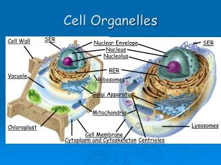
Other roles • Most organelles of eukaryotes, including the nucleus, endoplasmic reticulum, mitochondria, chloroplasts, lysosomes and vacuoles, are also formed from membranes. • These membranes form discrete compartments within the cell and control the movement of substances between these compartments. • Organelles are held in place by a network of fine protein filamentswithin a cell, collectively known as the cytoskeleton. • Prokaryotic cells such as bacteria lack these internal membranes. • Basic structure of all biological membranes is very similar although the plasma membrane is thicker than the membranes of intracellular organelles. • plasma membrane is 7-9nm thick • nuclear and endoplasmic reticulum membranes are 5-7nm thick
Membrane Structure • Basic structure of all biological membranes is very similar although the plasma membrane is thicker than the membranes of intracellular organelles. • plasma membrane is 7-9nm thick • nuclear and endoplasmic reticulum membranes are 5-7nm thick • The basic structure of a biological membrane is a lipid bilayer made up of phospholipid molecules. • The hydrophilic or water loving headsare attracted to water while the hydrophobic or water hating tails repel water. • The plasma membrane is surrounded by a watery environment both inside and outside the cell. • An important characteristic of the phospholipid bilayer is that the phospholipid molecules can move laterally in each layer.
Other membrane components • Steroid lipids • Embedded at intervals to give the membrane rigidity and water resistance. • Animal cells - cholesterol • Plant cells – phytosterol • Glycolipids • Formed by adding a carbohydrate to a lipid molecule • Common in the membranes of nerve and brain cells • Serve as surface markers.
Other membrane components • Glycoproteins • formed by adding a carbohydrate group to a protein. • act as receptors on the surface of the cell. • Proteins • A range of different proteins rest on the surface or penetrate through the phospholipid bilayer. • Classified according to how they are attached to the phospholipid bilayer. • Attachment of a protein to the hydrophobic lipid bilayer is due to the protein containing a hydrophobic group of amino acids. • Hydrophilic ends of the protein protrude from the outer and inner membrane surface into the aqueous environment.
Fluid mosaic model • Fluid mosaic model is the name given to describe the structure of plasma membranes. • We know that a plasma membrane comprises a phospholipid bilayer into which proteins and glycoproteins protrude. • The fluid part of the name comes from the fact that phospholipid molecules can move laterally in each layer. • The mosaic part of the model of membrane structure refers to the range of different proteins resting on the surface or penetrating through the phospholipid bilayer.
Membrane Proteins • Have various functions that reflect the functions of the membrane as a whole. • Transport proteins • Openings on both sides of the membrane • Form channels that allow some substances to move through the membrane • Receptor proteins • Bind hormones and other substances that cause changes to the cell’s activities. • Different types of cells have different receptor proteins enabling them to carry out different functions. • Recognition proteins • Are attached to carbohydrate molecules and act as markers, called antigens. • Allow the immune system to distinguish between the body’s own cells and foreign invaders. • Adhesion proteins • Link cells together in multicellular organisms.
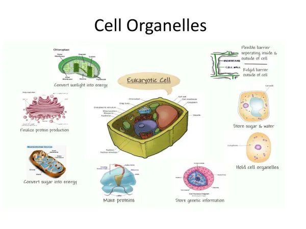
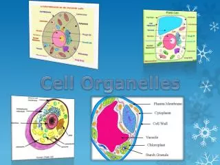
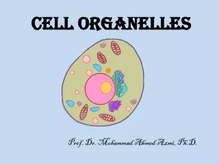
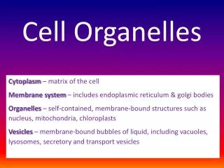
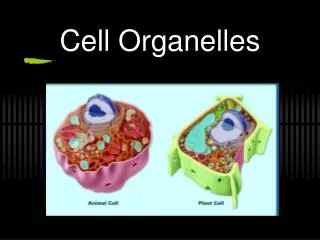
Cell Organelles • The synthesis, packaging and transport of organic molecules in eukaryotic cells involves the coordinated activity of a number of organelles.
Nucleus • The control centre of the cells of animals, plants, algae and fungi is the nucleus. • The nucleus in these cells forms a distinct spherical structure that is enclosed within a double membrane, known as the nuclear envelopewhich contains many nuclear pores. • The nucleus is usually the largest organelle in a cell (vacuoles in plant cells the only exception). • Most plant and animal cells have one nucleus. • Exceptions include skeletal muscle (more than one nucleus per cell) and human red blood cells (lose nucleus as they mature).
Nucleus • A light microscope view reveals that the nucleus contains many granules that are made of the genetic material (DNA). The DNA is usually dispersed within the nucleus. During the process of cell reproduction, however, the DNA granules become organised into a number of rod-shaped chromosomes. • Chromosomes are composed of super-coiled DNA which contains thegenetic information for production of specific proteins called enzymes which facilitate the manufacture of proteins, carbohydrates, lipids and nucleic acids required by cells. • DNA does not leave the nucleus. • Messenger RNA (mRNA) is produced in the nucleus and is a copy of the DNA instructions for a particular protein. Once produced, mRNA passes out into the cytoplasm through pores in the nuclear envelope. • The nucleus also contains one or more large inclusions known as nucleoli (singular – nucleolus) are an aggregation of ribonucleic acid (RNA) molecules.
Mitochondria • Mitochondria(singular = mitochondrion) are the powerhouse of the cell. • Each mitochondrion has an outer membrane and a highly folded inner membrane. • ATP is produced by reactions that occur on the inner folded membranes or cristae. • ATP (adenosine triphosphate)is the energy currency of cells. • The number of mitochondria present in different cells is related to the rate of energy usage by the cell. • Active cells, such as heart muscle cells, have many thousands of mitochondria. • Within a cell, mitochondria are found clustered in regions of high metabolic activity.
Ribosomes • Ribosomesare the organelles where protein production occurs. • They are not enclosed by a membrane. • Ribosomes are ~30nm in diameter and can only be seen using an electron microscope. • mRNA binds to ribosomes in order to initiate and direct protein synthesis. • The proteins produced by ribosomes on rough endoplasmic reticulum are transported to other parts of the cell and many are transported away from the cell. • Proteins made by ‘free’ ribosomes unattached to endoplasmic reticulum are for local use within the cell. • Mitochondria and chloroplasts also contain free ribosomes. • Chemical testing shows that ribosomes are composed of protein and ribonucleic acid (RNA).
Endoplasmic reticulum • The endoplasmic reticulum (ER) is responsible for transporting and processing proteins. • It is an extensive network of interconnected membranous sacs (cisternae), tubules and vesicles found throughout the cytoplasm of eukaryotic cells. • Contains enzymes and proteins required for the processing and sorting of proteins; adding sugar chains to proteins; and ensuring that proteins are correctly folded. • When ribosomes are attached to the surface of the ER it is referred to as rough endoplasmic reticulum (RER), otherwise it is referred to as smooth endoplasmic reticulum (SER). • Once proteins are synthesized by ribosomes on the RER they pass into the lumen of ER and travel to the Golgi apparatus in order to be exported from the cell. • RER is abundant in cells that actively synthesize and export proteins.
Golgi complex • Is linked to the endoplasmic reticulum. • Is the major site for the sorting of proteins to different sub-cellular compartments and packaging for export. • Proteins synthesized by the RER are often further modified in the Golgi before being packaged into secretory vesicles for export from the cell. • Secretory vesicles may be stored in the cytosol before they eventually fuse with the plasma membrane and release their contents from the cell in a process known as exocytosis. • Not all products packaged by the Golgi are intended for release from the cell – the enzymes in lysosomes are a key example of such a product. • Secretory cells have a well-developed Golgi while non-secretory cells have small Golgi.
Lysosomes • Are membrane-bound vesicles unique to eukaryotic cells. • Contain various powerful digestive enzymes capable of breaking down all the major classes of biological macromolecules • Are the sites of breakdown of debris within the cell and of external debris or foreign microorganisms that might be harmful. • The enzymes within lysosomes are produced in the ER and packaged into a transport vesicle in the Golgi. • This transport vesicle fuses with vesicles containing unwanted material and forms a lysosome. • Useful small molecules diffuse back into the cytoplasm while the residue is either retained by the lysosome or released from the cell by exocytosis. • Diseases resulting from these errors in lysosome enzymes include Tay Sachs disease, in which abnormal accumulation of lipids occurs, and Hurler syndrome, in which abnormal accumulation of complex carbohydrates occurs.
Peroxisomes & Endosomes • Peroxisomes and endosomes are small organelles that occur in eukaryotic cells and have some similarity with lysosomes. • Peroxisomes • Are small membrane-bound organelles rich in the enzymes catalase and urate oxidase. • Catalase prevents the accumulation of hydrogen peroxide (H2O2),a byproduct of many biochemical processes within cells. Hydrogen peroxide is a poison substance if allowed to accumulate. • Peroxisomes in different types of cells may contain different sets of enzymes. • Plant and animal cells have peroxisomes. • Endosomes • Endosomes are membrane-bound organelles found in animal cells. • When material enters a cell by endocytosis, endosomes pass on the newly ingested material to lysosomes for digestion.
Granuales • Granules • Are storage sites for biomolecules. • Organisms can maintain stores of chemical energy in the form of carbohydrates and lipids in organelles. • Examples of these are starch grains, lipid droplets and glycogen granules. • Plant cells store the complex carbohydrate starch in large grains e.g. cereal, while animal cells store glycogen or lipid droplets. • Plants such as legumes (beans and peas) can store protein granules, but animal cells cannot.
Vacuoles • Vacuoles • are membrane-bound, liquid-filled spaces found in most cells in variable numbers and sizes. • Food vacuoles in animal cells contain enzymes and are involved with intracellular digestion. • Contractile vacuoles in protozoa are involved in water balance, and expel excess water that enters by osmosis. • Vacuoles in plants provide physical support (turgidity) and storage. Salts are actively accumulated in the vacuole, and the vacuole is also a storage site for toxic substances. • Movement of substances into the plant vacuole is controlled by the vacuole membrane (the tonoplast).
Chloroplasts • Present in some cells of plants and algae. • Are typically 5 to 10 μm in length. • Capture the radiant energy of sunlight and transform it to chemical energy present in organic molecules, such as sugars, in a process known as photosynthesis. • The boundary of each chloroplast is a double membrane (inner and outer). • The inner membrane extends to form a system of membranous sacs called lamella or thylakoids. • When several of these stack together they form grana. • Chlorophyll is located in the grana and it is here that the light-dependent reactions of photosynthesis occur. • The semi-fluid substance between the grana is called the stroma and contains the enzymes necessary for the light-independent reactions of photosynthesis. • Chloroplasts also contain molecules of DNA, free ribosomes, starch grains and lipid droplets.
Cytoskeleton • Each cell has an internal framework of protein microtubules, microfilaments and intermediate filaments. • These three structures combine to assist in: • Maintaining the shape of a cell • Providing a support structure for other components within a cell • The movement of materials within a cell • Movement of the cell itself if required.
Components of the cytoskeleton • Microtubules are hollow and are made of subunits of the protein tubulin. • Microfilaments are solid, thinner and more flexible than microtubules. They are made of actin. • Intermediate filaments are made of a variety of proteins, depending on the particular cell, and are very tough. They often tie into the cytoskeleton of other cells.
Cells and multicellular organisms • Multicellular organisms consist of more than one cell. • This raises a few questions: • What connections, if any, exist between such cells? • What holds groups of cells together? • Do neighbouring cells or groups of cells communicate with each other in any way?
Connections between animal cells • There are three different types of junctions in animal cells: • Occluding junctions. • Communicating junctions • (also called gap junctions). • Anchoring junctions • (also called desmosomes).
Connections between animal cells • Occluding junctions • Occluding junctions involve cell membranes coming together in contact with each other. • There is no movement of material between cells. • Communicating junctions • Consist of protein-lined pores in the membranes of adjacent cells. • Permit the passage of salt ions, sugars, amino acids and other small molecules as well as electrical signals from one cell to another. • Example is the control of the beating of the heart. A small area of your heart, called the pace maker, receives an electrical impulse. This electrical impulse is transmitted to all cells of the heart through communication junctions so that the heart ‘beats as one’.
Connections between animal cells • Anchoring junctions • Anchoring junctions are the most common form of junction between epithelial cells in areas such as the skin or uterus. • Dense plaques of protein exist at the junction between two cells. • Fine fibrils extend from each side of these plaques and into the cytosol of the two cells involved. These are intermediate filaments that use the plaques as anchoring sites. • This structure has great tensile strength and acts throughout a group of cells because of the connections from one cell to another.
Connections between plant cells • Plant cells have no need for a structure such as the anchoring junctions of animal cells. • Why? • Structural support is provided by individual cell wall. • A layer of pectin (a sticky polysaccharide) holding the primary cell walls of adjacent cells tightly together. • In addition, secondary walls are laid down in each cell on the cytosol side of the primary wall resulting a relatively wide structure across the two cells (at least 0.1 μm thick). • The junctions that exist in plant cells to allow communication between adjacent cells in spite of the thick wall are plasmodesmata(singular: plasmodesma)
Plasmodesmata • Because of the way in which plant cell walls are built up, the gap or pore between two cells is lined with plasma membrane so that the plasma membrane of the two cells is continuous. • The desomtuble that bridges the ‘gap’ is continuous with the smooth ER of each cell. • Plasmodesmataexist in virtually all plants and hence cell-to-cell communication can occur between large numbers of cells that are in effect connected via their cytoplasm.