Lipids & Biological Membranes
300 likes | 620 Vues
Harini Chandra. Lipids & Biological Membranes.

Lipids & Biological Membranes
E N D
Presentation Transcript
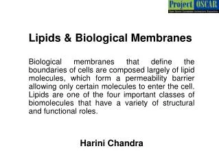
Harini Chandra Lipids & Biological Membranes Biological membranes that define the boundaries of cells are composed largely of lipid molecules, which form a permeability barrier allowing only certain molecules to enter the cell. Lipids are one of the four important classes of biomolecules that have a variety of structural and functional roles.
Master Layout (Part 1) 1 This animation consists of 4 parts: Part 1 – Fatty acids Part 2 – Membrane lipids Part 3 – Proteins in membrane structures Part 4 – Properties of cell membranes b w 2 2 w 3 1 a Fatty acid – general structure 3 w-3 double bond Degree of unsaturation Chain length 4 5 Source: Biochemistry by Lubert Stryer, 5th & 6th edition (ebook)
Definitions of the components:Part 1 – Fatty acids 1 1. Fatty acid: Fatty acids, more simply known as fats, are the key components of lipids that play an important role in signal transduction pathways and as structural elements of membranes. They are long hydrophobic chains of different lengths that possess a carboxylate group at one end. Most naturally occuring fatty acids have an even number of carbon atoms with varying degrees of unsaturation. 2. Degree of unsaturation: The number of double bonds present in a fatty acid chain defines its degree of unsaturation. 3. Chain length: The total number of carbon atoms present in a fatty acid chain is its chain length. 4. w-carbon: Fatty acids are numbered starting from their carboxyl group with the second and third carbon atoms being known as the a and b carbons. The carbon atom that is furthest away from the carboxylate group, at the distal end of the chain is known as the w carbon. 2 3 4 5
Part 1, Step 1: 1 Fatty acids – general structure w b 2 w-3 double bond w Degree of unsaturation 2 3 1 w-carbon Chain length Carboxylate group (C1) 3 a Saturated fatty acid Short chain length & increased unsaturation enhance fluidity & decrease MP of fatty acids. Unsaturated fatty acid 4 Action Description of the action Audio Narration As shown in animation. Fatty acids are long hydrophobic chains of carbon atoms having different lengths and a carboxylate group at one end. Fatty acids that do not contain any double bonds are said to be saturated while those possessing one or more double bonds in their structure are unsaturated. Most naturally occuring fatty acids have an even number of carbon atoms with varying degrees of unsaturation. The carboxylate group is numbered as one and the last carbon atom that is furthest away from the carboxylate group is known as the omega carbon. (Please redraw all figures.) First show the figure on the left appearing with its labels from the right end to the left in parts as depicted in the animation. Next, show the figure on the right with the labels appearing after the figure. 5 Source: Biochemistry by Lubert Stryer, 6th edition (ebook)
Part 1, Step 2: 1 Systematic name is derived by replacing the final ‘e’ of the parent hydrocarbon with ‘oic’/’oate’. For eg. C16 fatty acid is hexadecanoate (parent hydrocarbon is hexadecane). Fatty acids - nomenclature 12 14 6 4 2 16 8 10 1 13 9 3 11 7 5 15 Palmitic acid (C16, saturated) 2 3 Oleic acid (C18, monounsaturated) 2 1 Double bonds referred to by cis/trans- ∆-number at which the double bond is located in superscript. 4 17 3 15 18:1 indicative of 18 carbon atoms with 1 double bond. 6 18 5 13 16 11 14 7 9 8 10 Systematic name: cis-∆9-octadecenoate 12 4 Action Description of the action Audio Narration The fatty acid names are derived from their corresponding parent hydrocarbons by replacing the ‘e’ at the end with ‘oic’ or ‘oate’. A saturated fatty acid with 16 carbon atoms, for instance, is known as hexadecanoate. If there is one double bond, then it become deceneoate with the position of the double bond being indicated as a superscript after a delta symbol. For instance, a 18 carbon fatty acid with one double bond is known as ocatadecenoate while with two double bonds, it is known as octadecadienoate. As shown in animation. (Please redraw all figures.) First show the figure on top followed by the green callour located above it. Next show the figure below followed by the two text boxes and then the violet callout shown. 5 Source: Biochemistry by Lubert Stryer, 6th edition (ebook)
Master Layout (Part 2) 1 This animation consists of 4 parts: Part 1 – Fatty acids Part 2 – Membrane lipids Part 3 – Proteins in membrane structures Part 4 – Properties of cell membranes 2 Cholesterol GLYCEROL Fatty acid Fatty acid Sugar unit Fatty acid Alcohol 3 Phosphate Glycolipids 4 5 Phospholipids Source: Biochemistry by Lubert Stryer, 6th edition (ebook)
Definitions of the components:Part 2 – Membrane lipids 1 1. Lipids: These are water insoluble biomolecules that readily dissolve in organic solvents like chloroform and have a wide range of biological functions. They are important components of membranes, serve as fuel reserves and signalling molecules. Three important membrane lipids include phospholipids, glycolipids and cholesterol. 2. Phospholipid: Phospholipids are composed of four components – fatty acids, a platform to which the fatty acid is attached, phosphate residue and an alcohol attached to the phosphate. The platform to which the fatty acids are linked is commonly glycerol but in some cases, a more complex alcohol known as sphingosine may also be present. Phospholipids containing glycerol are known as phosphoglycerides, with two OH groups of glycerol being esterified with the carboxylate groups of fatty acids. The fatty acid chains form the hydrophobic tail while the remaining components constitute the hydrophilic head group. 3. Glycolipid: Lipids that have a sugar component in them are known as glycolipids. They are made up of a sphingosine backbone with the amino group acylated by a fatty acid and one or more sugar residues attached to the primary hydroxyl group. The simplest glycolipid is known as cerebroside which contains either glucose or galactose as its sugar residue. 4. Cholesterol: Cholesterol is a steroid molecule whose structure is significantly different from that of phospholipids and glycolipids. Cholesterol is found in varying quantities in animal membranes but is not present in prokaryotes. It is composed of a hydrocarbon chainlinked to one end and a hydroxyl group at the other end. 2 3 4 5
Part 2, Step 1: 1 Membrane lipids - phospholipids GLYCEROL Fatty acid Fatty acid Phosphatidate (Diacylglycerol 3-phosphate) 2 Alcohol Phosphate Glycerol backbone Phosphate group 3 Fatty acids Shorthand depiction Ester linkage Polar head group Hydrophobic tail 4 Action Description of the action Audio Narration (Please redraw all figures.) First show the coloured block diagram structures on left top. First the blue rectangle must appear followed by the two ovals and then the green rectangle and finally the violet parallelogram. Next, the structure on bottom right must appear. First the central region must appear marked ‘glycerol backbone’. Next, the groups on the left marked ‘fatty acids’ must appear followed by the pink group on the right. The green box must then highlight the region as indicated with the corresponding label. The figure on bottom left must then appear with the labels. Phospholipids are composed of four components – fatty acids, a platform to which the fatty acid is attached, phosphate residue and an alcohol attached to the phosphate. The platform to which the fatty acids are linked may be glycerol or sphingosine. Phospholipids containing glycerol are known as phosphoglycerides, with two OH groups of glycerol being esterified with the carboxylate groups of fatty acids. The simplest phospholipid, phosphatidate, is made up of only the phosphate group and fatty acids attached to the glycerol backbone. As shown in animation. 5 Source: Biochemistry by Lubert Stryer, 6th edition (ebook)
Part 2, Step 2: 1 Common phosphoglycerides Glycerol backbone 2 Fatty acids 3 Phosphatidyl ethanolamine Phosphatidyl serine Phosphatidyl choline Phosphatidyl inositol Diphosphatidyl glycerol (cardiolipin) 4 Action Description of the action Audio Narration The important phophoglycerides found in membranes are derived from phosphatidate by esterification of the phosphate group with the hydroxyl group of various alcohols. The most commonly observed phosphoglycerides include phosphatidyl serine, choline, ethanolamine, inositol and diphosphatidyl glycerol, also known as cardiolipin. As shown in animation. (Please redraw all figures.) First show the blue and green parts of the structure with their labels. Next, the red groups must appear sequentially one after another with their corresponding names appearing below as shown in animation. (The red groups are layered one over another – watch in slide show mode only) 5 Source: Biochemistry by Lubert Stryer, 6th edition (ebook)
Part 2, Step 3: 1 Phospholipids - sphingomyelin 2 Sphingosine Fatty acid 3 Choline Sphingomyelin 4 Action Description of the action Audio Narration Sphingosine is another amino alcohol backbone that serves as a platform for attachment of fatty acids and alcohols. Sphingomyelin, derived from sphingosine , consists of a fatty acid linked to the amino group via an amide bond and a choline moiety attached to the primary hydroxyl group via a phosphate group. As shown in animation. (Please redraw all figures.) First show the figure on top with its label followed by the figure below with its labels as shown. 5 Source: Biochemistry by Lubert Stryer, 6th edition (ebook)
Part 2, Step 4: 1 Membrane lipids - glycolipids Fatty acid Sugar residue(s) 2 SPHINGOSINE 3 Cerebroside 4 Action Description of the action Audio Narration Lipids that have a sugar component in them are known as glycolipids. They are made up of a sphingosine backbone with the amino group acylated by a fatty acid and one or more sugar residues attached to the primary hydroxyl group. The simplest glycolipid is known as cerebroside which contains either glucose or galactose as its sugar residue. As shown in animation. (Please redraw all figures.) First show the box diagrams shown on top left. The green rectangle must appear first follwed by the blue parallelogram and then the brown oval. Next show the structure below in which the blue region must appear first followed by the green region and finally the pink labeled box. 5 Source: Biochemistry by Lubert Stryer, 6th edition (ebook)
Part 2, Step 5: 1 Membrane lipids - cholesterol 2 Alkyl side chain 3 D C Polar head group B A Steroid nucleus 4 Action Description of the action Audio Narration As shown in animation. (Please redraw all figures.) Show the appearance of the structure above. First ring A must appear followed by ring B, then ring C and then ring D. All the other groups attached at the various positions must appear after all four rings have appeared. Another group of structural membrane lipids is the sterols, found in most eukaryotic cells. The steroid nucleus consists of four fused rings that are oriented in a planar manner. Cholesterol is an amphipathic molecule with a polar hydroxyl head group and a non-polar steroid nucleus and hydrocarbon side chain. In addition to having a structural role in membranes, sterols are precursors for several products such as steroid hormones and bile acids. 5 Source: Biochemistry by Lubert Stryer, 6th edition (ebook)
Part 2, Step 6: 1 Micellar arrangement Membrane formation by phosphoglycerides Hydrogen bonding, electrostatic attraction Polar head group Hydrophobic tail 2 Hydrophobic, Van der Waals interactions 3 Random orientation Bilayer arrangement 4 Action Description of the action Audio Narration The amphipathic nature of the phosphoglyceride molecules consisting of a polar head group and a hydrophobic tail enables them to rearrange themselves in an aqueous environment. When they come in contact with aqueous surroundings, they can either reorient to form a micellar structure or a bilayer arrangement. In these arrangements, the polar head groups are in contact with water by means of hydrogen bonding while the hydrophobic tails interact with each other through hydrophobic and Van der Waals interactions. The lipid bilayer arrangement is more favoured for phospholipids and glycolipids. As shown in animation. (Please redraw all figures.) First show the green structures oriented randomly. Next they must be surrounded by water (blue clouds). When this happens, the green structures must reorient themselves in the two arrangements shown on the right after the arrows. The arrows and green ovals with the text must then appear. 5 Source: Biochemistry by Lubert Stryer, 6th edition (ebook)
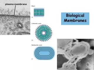
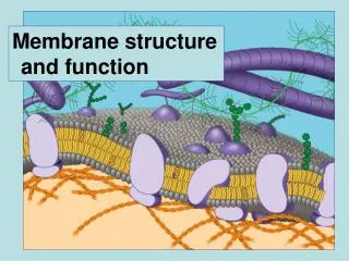
Master Layout (Part 3) 1 This animation consists of 4 parts: Part 1 – Fatty acids Part 2 – Membrane lipids Part 3 – Proteins in membrane structures Part 4 – Properties of cell membranes Glycoprotein 2 Outside Glycolipid Integral protein 3 Lipid bilayer Phospholipids 4 Sterol Inside Peripheral protein 5 Source: Biochemistry by A.L.Lehninger, 4th edition (ebook)
Definitions of the components:Part 3 – Proteins in membrane structures 1 1. Lipid bilayer: The flat membrane sheets that form a barrier around cells consisting of two layers of lipid molecules is known as the lipid bilayer. The hydrophobic tail regions are sequestered within the bilayer, away from the aqueous environment while the polar heads face outward and interact with the surrounding molecules. The bilayer is also embedded with proteins that perform specific functions for the cell. 2. Integral proteins: Those proteins that span the membrane and are embedded within the lipid bilayer are known as integral proteins. They interact extensively with the hydrophobic chains of lipids and cannot be easily dissociated from the membrane. 3. Peripheral proteins: Peripheral membrane proteins, however, are only bound to the membrane surfaces by means of electrostatic and hydrogen bond interactions with the polar head groups of the lipids. They can be easily dissociated from the membrane with mild agents such as salts, acids or alkali since they are not embedded within it. 4. Glycoprotein: Carbohydrate groups are often covalently attached to proteins to form glycoproteins. The sugar residues are typically attached to the amide nitrogen atom of the aspargine side chain or to the oxygen atom of the serine or threonine side chain. These glycoproteins are components of cell membranes and have a variety of functions in cell adhesion processes. 2 3 4 5
Part 3, Step 1: 1 Integral proteins - Bacteriorhodopsin Amino acid sequence of membrane protein A Q I T G RPE W I W L A L G T A L M G L G T L Y F L VK G M G V S DPDAK KF Y A I T T L V P A I A F T M Y L S M L L G Y G L T M V P F G G E Q N P I Y W A RY A DW L F T T P L L L LDL A L L V D A DQ G T I L A L V G ADG I M I G T G L V G A L T K V Y S YRF V W W A I S T A A M L Y I L Y V L F F G F T S K A E S M R P E V A S T FKV LRN V T V V L W S A Y V V V W L I G S E G A G I V P L N IET L L F M V L DV S AKV G F G L I L LR S R A I F G E A E A P E P S A D G A A A T S 2 3 Residues of the 7 membrane-spanning helices (largely non-polar) Membrane spanning a-helices Charged residues 4 Action Description of the action Audio Narration As shown in animation. Bacteriorhodopsin is an archaeal integral membrane protein that plays a role in energy transduction, using light energy for the transport of protons from inside to outside the cell. It is made of seven membrane-spanning alpha helices that are oriented perpendicular to the plane of the membrane. Determination of the amino acid sequence of this protein revealed that most of the residues within the membrane are non-polar, thereby allowing favorable interactions with the lipid hydrocarbon chains. Very few charged residues were found in the structure. (Please redraw all figures.) First show the figure on the left. The red box must then appear which must be zoomed into to show the figure on the right with the highlighted regions as depicted along with all the labels and the key shown below. 5 Source: Biochemistry by Lubert Stryer, 6th edition (ebook)
Part 3, Step 2: 1 - - - Integral proteins – Porins - Hydrogen bonded b-strands Amino acid sequence of porin from Rhodopseudomonas blastica 2 Hydrogen bonds 3 Hydrophobic residues (on surface of structure) Hydrophilic residues (buried inside) 4 Action Audio Narration Description of the action Porins are another class of integral membrane proteins that form channels within the membrane. They are composed entirely of b-strands with essentially no alpha helices in their structure. These beta strands are hydrogen bonded to each other to form a beta sheet which folds to form a hollow cylindrical structure. The folding occurs such that the polar amino acid residues line the inside of the cylinder, thereby making it hydrophilic. This allows the channel to be filled with water and also allows passage of small ions and charged molecules. The non-polar residues facing outside interact hydrophobically with the lipid chains of the membrane. As shown in animation. (Please redraw all figures.) First show the structure on top with its label. Then show the dotted arrows and the figure on right top. The brown circles must appear and pass through the blue cylinder. This must happen continuously throughout this animation. Simultaneously, the green box must appear and this region must be zoomed into and the figure below must be shown with its labels and the key on the left. 5 Source: Biochemistry by Lubert Stryer, 6th edition (ebook)
Part 3, Step 3: 1 Peripheral proteins Acid/alkali added – change in pH Dissociation Integral proteins 2 Peripheral membrane proteins 3 Dissociation 4 Action Description of the action Audio Narration As shown in animation. Peripheral membrane proteins are attached to either the outside or inside surface of the membrane via electrostatic and hydrogen bond interactions with either the lipid heads of the membrane or with other integral proteins. These polar interactions can be easily disrupted by addition of acids or alkali which modify the pH or by addition of salts. (Please redraw all figures.) First show the figure in the middle with the yellow and blue shapes labeled a-e. Next show the blue cloud appearing on the blue shapes with the corresponding label. The blue shapes, d & e, must then dissociate from the figure as shown. 5 Source: Biochemistry by A.L.Lehninger, 4th edition (ebook)
Part 3, Step 4: 1 Each amino acid is associated with a free energy change for its transfer from a hydrophobic to aqueous environment. Prediction of transmembrane helices – Hydropathy index Threshold value for helix detection 2 Hydropathy index, kJ/mol 3 ~ 20 amino acid residues ~ 30 Ao Free energy calculations are made for transfer of every 20 amino acid residues (i.e. 1-20, 2-21, 3-22 etc.) from hydrophobic to aqueous environment. This is plotted as a hydropathy plot. First amino acid residue in window 4 Action Description of the action Audio Narration As shown in animation. It is possible to predict transmembrane helix regions of a protein by calculating the free energy changes associated with the transfer of residues from a hydrophobic to aqueous environment. The width of a membrane is typically around 30Ao, which can fit approximately 20 amino acid residues. Therefore the free energy change for hypothetical alpha helices formed every 20 residues, from residue1 to 20, 2 to 21, 3 to 22 and so on are calculated until the end of the sequence is reached. These free energy changes are plotted against the first amino acid residue of every 20-residue window ito obtain a hydropathy plot. A peak above 84 kJ/mol is indicative of a likely membrane spanning helix. This, however does not detect membrane spanning b sheets. (Please redraw all figures.) First show the long chain like structure shown in the centre with the labels. Next show the dialogue box on the right top followed by the dialogue box at the bottom. Once this is shown, the graph on the right must appear with the arrow mark and text box. 5 Source: Biochemistry by Lubert Stryer, 6th edition (ebook)
Master Layout (Part 4) 1 This animation consists of 4 parts: Part 1 – Fatty acids Part 2 – Membrane lipids Part 3 – Proteins in membrane structures Part 4 – Properties of cell membranes Bleach Fluorescence intensity Fluorescence Recovery After Photobleaching (FRAP) 2 Recovery Recovery Bleach Time 3 Lateral diffusion Very fast 1 mm/s 4 Transverse diffusion (flip-flop) Very slow t1/2 in days 5 Source: Biochemistry by A.L.Lehninger, 4th edition (ebook)
Definitions of the components:Part 4 – Properties of cell membranes 1 1. Fluorescence Recovery After Photobleaching (FRAP) : This is a technique by which a cell surface component is first labelled by means of a fluorescent molecule and a small region of the cell surface is viewed by means of fluorescence microscopy. The fluorescent molecules in the region being viewed are destroyed by a laser pulse, a process known as bleaching. Once this occurs, the time required for fluorescence to reappear in this region is plotted against the fluorescence intensity. This helps in understanding the movement of molecules across the cell surface. 2. Lateral diffusion: The process by which membrane components move laterally from one region to another in the same plane. This is a quick process and takes place in a matter of microseconds. Proteins exhibit varying degrees of lateral mobility, with some being as mobile as lipids and others being almost immobile. 3. Transverse diffusion (flip-flop): This is a process by which molecules in the membrane transition from one surface of the membrane to the other. The time required for transverse diffusion is significantly more than that for lateral diffusion and can be measured by electron spin resonance techniques. This process is made quicker by the enzyme ‘flippase’. 4. Fluid Mosaic Model: The overall organization and properties of biological membranes were proposed by Jonathan Singer and Garth Nicolson in 1972 as the Fluid Mosaic Model. They proposed that membranes are two-dimensional solutions of oriented lipids and globular proteins, with the lipids serving as a “solvent” for integral membrane proteins and functioning as a permeability barrier. They also hypothesized that membrane proteins undergo lateral diffusion freely but not transverse diffusion. 2 3 4 5
Part 4, Step 1: 1 Lateral diffusion of membrane components - FRAP Fluorescence intensity Bleach 2 Laser Recovery Bleaching Recovery Time 3 Region being viewed through microscope Cell surface components labelled with fluorescent molecule 4 Action Description of the action Audio Narration As shown in animation. Lateral diffusion of membrane components can be proved using fluorescence recovery after photobleaching technique. A cell surface component is first labelled by means of a fluorescent molecule and a small region of the cell surface is viewed by means of fluorescence microscopy. The fluorescent molecules in the region being viewed are destroyed by a laser pulse, a process known as bleaching. Fluorescence however reappears in the region after a certain time that is dependent on the diffusion coefficient of the molecules. (Please redraw all figures.) First show the blue figure with the green spots on it and the corresponding labels. Followed by the red box and its label. Next, show the ‘laser’ and its light falling on the green spot at the bottom. Once this happens, the green spot must change color to grey and the label ‘bleaching’ must appear. Then the laser must be removed and the grey spot should move down and disappear and simultaneously the green spot on top must move into the red box as shown in animation. When bleaching occurs, the downward slope of the graph must be shown and when the green spot on top enters the red box, the upward curve must be shown to appear. 5 Source: Biochemistry by Lubert Stryer, 6th edition (ebook)
Part 4, Step 2: 1 Lateral diffusion Vs Transverse diffusion Lateral diffusion Very fast 2 1 mm/s Flippase 3 Transverse diffusion (flip-flop) Very slow Very fast t1/2 in seconds t1/2 in days 4 Action Audio Narration Description of the action The Fluid Mosaic model explains the lateral diffusion of membrane components but not the transverse diffusion. Lateral diffusion is a rapid process taking place in the range of microseconds. However, transverse diffusion, also known as the ‘flip flop’ reaction takes place very slowly over a period of several hours. This reaction is facilitated by the enzyme flippase, which carries out transverse diffusion in the time range of few seconds. As shown in animation. (Please redraw all figures.) First show the green figures on top with the title ‘lateral diffusion’. The blue shape must move as indicated by the arrow and reach the position indicated on the right. This must occur quickly. Next show the figure on left bottom with the title ‘transverse diffusion’. The blue shape alone must flip very slowly in the direction indicated by the arrow to reach the position shown on the right. Next, the brown oval must appear with its label. When this happens, the flipping must take place quickly in the same way as the previous diagram. 5 Source: Biochemistry by Lubert Stryer, 6th edition (ebook)
Part 4, Step 3: 1 Length of fatty acyl chain – longer chain, higher Tm Factors determining membrane fluidity Presence of cholesterol – greater cholesterol, higher Tm Fluid-like 2 3 Solid-like Degree of unsaturation – Saturated fatty acids increase Tm Tm Temperature 4 Action Description of the action Audio Narration As shown in animation. The fluidity of any biological membrane is dependent on the properties of the fatty acid chains present in it. Transition of the membrane from a rigid state to a fluid state occurs abruptly as the temperature is increased and crosses the melting temperature, Tm. This melting temperature is a function of the length of fatty acyl chains present and their degree of unsaturation. Increase in length of fatty acyl chain increases the Tm while increase in the degree of unsaturation decreases the Tm. In other words, greater number of double bonds disrupts the packing order achieved by saturated fatty acids thereby decreasing the Tm. In animals, the cholesterol content is another regulator of fluidity. Greater the amount of the bulky steroid, higher is the Tm. (Please redraw all figures.) The graph must appear gradually. As the graph is appearing in the centre, the text around it must appear sequentially as shown. 5 Source: Biochemistry by Lubert Stryer, 6th edition (ebook)
Interactivity option 1:Step No: 1 1 Membrane lipids are useful for designing lipid vesicles known as liposomes, which consist of small aqueous compartments surrounded by a lipid bilayer. These liposomes are increasingly being used as drug delivery systems in hydrophobic environments. Shown below is an example for formation of a glycine-containing liposome. Click on the green phospholipid layer to view liposome formation and then answer the question below. 2 Glycine in water Gel filtration Sonication 3 Phospholipid (Click here) 4 Results Interacativity Type Options (Please redraw all figures.) User must click on the green layer at the bottom of the first figure to view the animation after which the question with 4 options must appear and user must be allowed to choose any 1 option. When the user clicks on the green layer, the animation must be shown. The green layer shown at the bottom must gradually form green circles as shown in the middle panel and must surround a few red dots. Once this happens, the arrow saying ‘gel filtration’ must be shown and the red dots must disappear leaving only the green circles enclosing the red circles. The user must then answer the question shown in the next slide. Correct answer is C. If user gets it right, ‘correct answer’ must be displayed otherwise ‘wrong answer’ must be displayed. Click to view experiment & then choose the correct answer. 5
Interactivity option 1:Step No: 1 1 What property of membrane lipids allows them to form such liposome vesicles? 2 A) Their low melting temperature B) The presence of cholesterol C) Their self-sealing nature 3 D) The presence of glycerol in the phospholipids 4 Results Interacativity Type Options (Please redraw all figures.) User must click on the green layer at the bottom of the first figure to view the animation after which the question with 4 options must appear and user must be allowed to choose any 1 option. When the user clicks on the green layer, the animation must be shown. The green layer shown at the bottom must gradually form green circles as shown in the middle panel and must surround a few red dots. Once this happens, the arrow saying ‘gel filtration’ must be shown and the red dots must disappear leaving only the green circles enclosing the red circles. The user must then answer the question shown in the next slide. Correct answer is C. If user gets it right, ‘correct answer’ must be displayed otherwise ‘wrong answer’ must be displayed. Click to view experiment & then choose the correct answer. 5
Questionnaire 1 1. How many double bonds would be present in a fatty acid having the systematic name “all-cis-∆9, ∆12, ∆15-Octadecatrienoate”? Answers: a) 1 b) 2 c) 3 d) 4 2. Which of the following is a saturated fatty acid with 18 carbon atoms? Answers: a) cis- ∆9-Octadecenoateb) Octadecanoate c) Eicosanoate d)Tetradecanoate 3. Which of the following components is not present in Phosphatidyl inositol? Answers:a) Sphingosine b) Glycerol c) Phosphate d) Inositol 4. If the degree of unsaturation of fatty acyl chains increases, what happens to the Tm? Answers:a) Tm increases b) Tm remains same c) Tm decreasesd) None of the above 5. The threshold value of hydropathy index for detection of alpha helices is? Answers:a) -22 kJ/mol b) +22 kJ/mol c) +67 kJ/mol d) +84 kJ/mol 2 3 4 5
Links for further reading Books: Biochemistry by Stryer et al., 6th edition Biochemistry by A.L.Lehninger et al., 4th edition Biochemistry by Voet & Voet, 3rd edition Research papers: Singer, S. J. & Nicolson, G. L. The Fluid Mosaic Model of the Structure of Cell Membranes. Science 1972, 175 (4023), 720-731.