Download
1 / 103
1.04k likes | 1.23k Vues
Cardiovascular and Respiratory Systems: Oxygen Transport. Integration of Ventilation, Cardiac, and Circulatory Functions. Cardiorespiratory System. Functions of cardiorespiratory system : transportation of O 2 and CO 2 transportation of nutrients/waste products distribution of hormones

E N D
Cardiovascular and Respiratory Systems: Oxygen Transport Integration of Ventilation, Cardiac, and Circulatory Functions
Cardiorespiratory System Functions of cardiorespiratory system: • transportation of O2 and CO2 • transportation of nutrients/waste products • distribution of hormones • thermoregulation • maintenance of blood pressure
Ability of cardiorespiratory system on maintaining arterial PO2 (PaO2)during graded exercise to exhaustion
Critical elements of O2 Transport Pathway • Lungs • Ventilation • VE = RR VT • O2 diffusion into blood • PO2 gradient determines O2 movement • Hb • Heart and circulation • Q = HR SV • cardiac output = muscle blood flow • O2 diffusion into mitochondria • oxyhemoglobin dissociation relationship • Fick principle [VO2 = Q (CaO2 – CvO2)] • Control of cardiorespiratory system • central control • peripheral inputs • maintenance of blood pH
Ventilation and Diffusion Getting O2 from air into blood
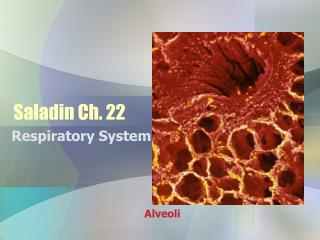
A. Major pulmonary structureB. General view showing alveoliC. Section of lung showing individual alveoliD. Pulmonary capillaries within alveolar walls
Pulmonary Gas Exchange • gases move because of pressure (concentration) gradients • alveolar thickness is ~ 0.1 µm • total alveolar surface area is ~70 m2 • at rest, RBCs remain in pulmonary capillaries for 0.75 s (capillary transit time) • transit time = 0.4-0.5 s at maximal exercise • adequate time to release CO2 • marginal time to take up O2
Pressure gradients for gas transfer at rest: Time required for gas exchange in lungs (left) and tissue (right)
What would be the effect on the saturation of arterial blood with O2 (SaO2) when pulmonary blood flow is faster than RBC can uptake O2? • SaO2 would remain unchanged • SaO2 would be decreased • SaO2 would be increased
What effect might a decreased SaO2 have on O2 utilization by mitochondria? • no effect on mitochondrial VO2 • will decrease mitochondrial VO2 • will increase mitochondrial VO2
Pulmonary circulation • Pulmonary circulation varies with cardiac output
Single alveoli at rest showing individual RBCs RBC Single alveoli under high flow showing increased RBCs
Gas Exchange and Transport Oxygen transport • ~98% of O2 transported bound to hemoglobin • 1-2% of O2 is dissolved in blood
Hemoglobin • consists of four O2-binding heme (iron containing) molecules • combines reversibly w/ O2 (forms oxy-hemoglobin)
Rate of gas diffusion is dependent upon pressure (concentration) gradient. Erythrocyte (RBC) ~98% of O2 is bound up with hemoglobin (Hb) and transported from lungs to working muscle.
Transport of O2 and CO2 in blood CO2 + H2O H2CO3 H+ + HCO3-
Predict the relative O2 pressure differences between alveoli (PAO2) and arterial blood (PaO2) • PAO2 > PaO2 • PAO2 = PaO2 • PAO2 < PaO2
Role of the Heart Moving O2 from lungs to working muscle
Cardiac Cycle • systole diastole • cardiac output (Q) = stroke volume (SV) heart rate (HR) examples • rest: SV = 75 ml; HR = 60 bpm; Q = 4.5 Lmin-1 • exercise: SV = 130 ml; HR = 180 bpm; Q = 23.4 Lmin-1
Control of cardiac function and ventilation Parallel activations
Reflex control of cardiac output Primary regulators • cardiovascular control center (medulla) • w/ activation of motor cortex, parallel activation of sympathetic/parasympathetic nerves • parasympathetic inhibition predominates at HR <~100 bpm • sympathetic stimulation predominates at HR >~100 bpm • skeletal muscle afferents • sense mechanical and metabolic environment Secondary regulator • arterial baroreceptors • located in carotid bodies and aortic arch • respond to arterial pressure • Reset during exercise
Cardiac Regulation Intrinsic control • Frank-Starling Principle • Ca2+ influx w/ myocardial stretch Extrinsic control • autonomic nervous system • sympathetic NS (1 control at HR >100 bpm) • parasympathetic NS (1 control at HR <100 bpm) • peripheral input • chemoreceptors, baroreceptors, muscle afferents • hormonal • EPI, NE (catecholamines)
Humoral Chemoreceptors • PaO2 • not normally involved in control • PaCO2 • central PaCO2chemoreceptors are 1º control factor at rest • H+ • peripheral H+ chemoreceptors are important factor during high-intensity exercise
Control of Ventilation • Central command and muscle afferents are primary control mechanisms • H+ chemoreceptors responsible for “fine-tuning” ventilation
Describe the mechanisms that control cardiac output and ventilation.
Cardiac output affected by: • preload – end diastolic pressure (amount of myocardial stretch) • afterload – resistance blood encounters as it leaves ventricles • contractility – strength of cardiac contraction • heart rate
Venus Blood Return to HeartSV dependent on venous return • muscle pump • one-way venous valves • breathing Return of blood to heart
Cardiovascular Response to Exercise Fick equation VO2 = Q (aO2 – vO2) VO2 = [HR SV] (aO2 – vO2) VO2 = [BP TPR] (aO2 – vO2)
VO2 = Q (aO2 – vO2) How would VO2 be affected if cardiac output/O2 extraction were increased? • increased • decreased • no effect • cannot be determined
Matching O2 delivery to muscle O2 needs Regulation of cardiorespiratory system
Exercise effects on heart • HR caused by • sympathetic innervation • parasympathetic innervation • release of catecholamines • SV, caused by • sympathetic innervation • venous return • cardiac output
Increasing Blood Flow to Working Muscle During Exercise Blood flow redistribution
Blood vessels are surrounded by sympathetic nerves. A feed artery was stained to reveal catecholamine-containing nerve fibers in vascular smooth muscle cell layer. This rich network extends throughout arterioles but not into capillaries or venules.
Local blood flow control • general sympathetic response occurs with exercise onset that causes vasoconstriction • exercise hyperemia = increase in blood flow to cardiac and skeletal muscle • blood flow to working muscle increases linearly with muscle VO2 • muscle metabolic rate is key in controlling muscle blood flow • controlled primarily by local factors
(1-adrenergic receptor blocker) 30 s Onset of exercise
Capillaries • flow of blood • aorta arteries arterioles capillaries venules veins vena cava • arterioles regulate blood flow into muscle • under sympathetic and local control • precapillary sphincters fine tune blood flow within muscle • under only local control • adenosine, PO2, PCO2, pH, nitric oxide (NO)