Meninges and CSF
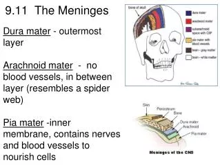
Meninges and CSF. Dr. K. Sivapalan. Meninges. Dura mater, arachnoid mater and pia mater. Dura- single and tough layer of fibrous tissue fused with inner periosteum except in few areas. Arachnoid - fibrocellular tissue. Outer most cells are bonded to each other by tight junctions.

Meninges and CSF
E N D
Presentation Transcript
Meninges and CSF Dr. K. Sivapalan
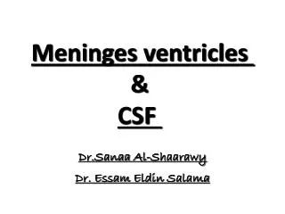
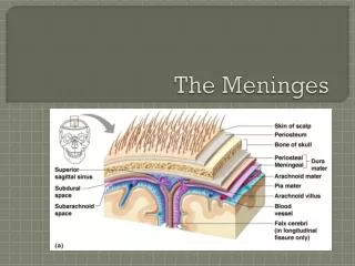
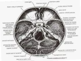
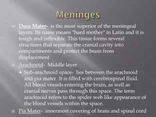
Meninges • Dura mater, arachnoid mater and pia mater. • Dura- single and tough layer of fibrous tissue fused with inner periosteum except in few areas. • Arachnoid- fibrocellular tissue. Outer most cells are bonded to each other by tight junctions. • Subarachnoid space- arachnoidtrabeculae cross the space to reach the pia. [CSF] • Pia- invests the nervous tissue closely lining the outer surface. It and the sub-pial space follow into the tissue with blood vessols. Meningies and CSF
Choroid Plexuses • Choroid plexus is found in the 4 ventricles. Meningies and CSF
CSF and ECF in Brain • CSF volume is about 150 ml, production is 550 ml/day, 50-70 % secreted by choroid plexus and the rest by vessels in the walls of the ventricles. • The composition is the same as the ECF in the brain which is 15 % of the brain volume. • The pia is freely permiable to CSF and there is free communication and diffusion of substances between the CSF and the ECF. Meningies and CSF
Circulation of CSF • CSF enters the third ventricle through the inter ventricular foramen. • It descends to the fourth ventricle through the aqueduct. • It enters the subarachnoid space through median and lateral apertures in the roof of the fourth ventricle. • Flow in the central canal of the spinal cord is negligible • Some CSF descends through foramen magnum reaching the lumbar cistern in 12 hours. • Small amount is absorbed into the spinal veins but most returns to subarachnoid space in cranium. • It ascends further through the tentorial notch and around the cerebrum. • Finally it is absorbed into the venous sinuses. Meningies and CSF
Function of the CSF • The weight of brain in air is 1400 g but in the “water bath” of CSF, it is 50 g. • The brain is attached and held in position by arachnoidtrabaculae, blood vessels and nerve roots. Weight reduction by CSF helps the flimsy attachments to hold the brain. • Removal of CSF can cause sever pain because the brain hangs on the vessols. • The brain is protected from the trauma of head injuries by the CSF and meninges. Meningies and CSF
Abnormalities of CSF • When circulation is blocked CSF pressure increases proximal to the obstruction. • Block of foramina in the fourth ventricle or failure of absorption results in hydrocephalus. • Abnormalities in composition signifies the type of disease of the meninges. Meningies and CSF