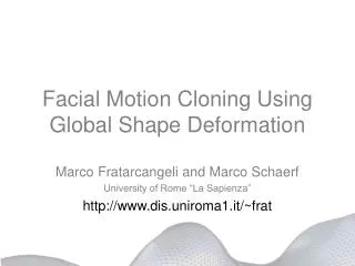
Measuring Anatomy and its Deformation Using Deformable Shape Models
230 likes | 260 Vues
Learn about measuring anatomy and deformations using deformable shape models, modeling morphological changes, spatial normalization, biomechanical modeling, and challenges in population imaging studies. Explore methods such as shape-based elastic transformation and adaptive focus deformable models for reconstruction and mapping. Discover applications in image-guided surgical planning and brain tumor segmentation. Collaborators at Johns Hopkins University School of Medicine and the Center for Biomedical Image Computing have contributed to advancements in anatomical deformation prediction and analysis.

Measuring Anatomy and its Deformation Using Deformable Shape Models
E N D
Presentation Transcript
Measuring Anatomy and its Deformation Using Deformable Shape Models Christos Davatzikos Center for Biomedical Image Computing Department of Radiology and Radiological Science Johns Hopkins University School of Medicine http://cbic.rad.jhu.edu
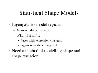
Topics • Deformable Shape Modeling and Registration • Modeling and Predicting Anatomical Deformations Quantitative Morphology Spatial normalization Data pooling Image Data Mining Statistical Atlases • Biomechanical/statistical modeling of tumor growth • Modeling intra-operative deformations
Challenges in population imaging studies: • Inter-individual anatomical variability • Localized subtle effects of disease on structure or function Brain atrophy, functional activation, or gene expression Need good alignment to increase sensitivity of statistical analysis in a standardized reference space Before Spatial Normalization After Spatial Normalization
Shape-Based Elastic Transformation in 3D: • 1) Shape reconstruction of a number of structures • (open or closed surfaces, curves) • 2) Match the features based on their geometric • properties (e.g. curvatures) • 3) Use the feature-to-feature map to drive an elastic • deformation transformation Davatzikos, JCAT/CVIU, 1996/1997
Examples of surfaces that drive the elastic deformation Sulcal Ribbons Cortex Hippocampus Basal ganglia and ventricles
1 2 3 4 Davatzikos, Human Brain Mapping, Jan. 1998
atrophy original Validation of the RAVENS methodology: Simulation of atrophy in precentral and superior temporal gyri
Localized atrophy identified via t-maps of the RAVENS images • Atrophy detected in the two gyri: PCG and STG • T-maps are overlaid on the average WM RAVENS map of 24 subjects
Adaptive Focus Deformable Model (AFDM) • for shape reconstruction and mapping: • Use of an Attribute Vector on each point of the model • Use of an Adaptive Deformation Approach Attribute Vectors: Affine-invariant geometric characteristics from a local to a global scale B C A Shen et.al., IEEE-TMI April 2001
Adaptive nature of AFDM 1. Model can zoom to small important features • In Active Shape Models, large variable features dominate over small variable, yet important features. Identical weighting Large weights on eyes mode 1 mode 2
Different weighting for different parts of the model White: reliable parts large weights Black: unreliable parts small weights
Shape / Volumetric Analysis of the Hippocampus Correlation Coefficient Averaging 0.975
Modeling and Predicting Anatomical Deformability with applications in image-guided surgical planning
Deformable Brain Atlas for Brain Tumor Patients • Segmentation for surgical planning purposes • Statistical atlases linking structural variables, surgical procedure, and surgical outcome Outcome A Outcome B
Kyriacou et.al., IEEE-TMI, 1998 Simulation of tumor growth via a biomechanical model
Fundamental Limitation: Estimating the inverse deformation field is a very ill-posed problem Atlas Unknown normal brain Patient’s brain deformed by tumor Unknown initial tumor position
Using shape statistics to model the deformation between pre- and intra-operative anatomy Pre-operative plan Intra-operative anatomy • Need to be able to predict anatomical deformations in the • planning stage • If part of a structure is visible intra-operatively but another • part is missing, the latter can be predicted Gray predicted from green
Generation of training samples, using biomechanical models or available images (e.g. intra-operative images) s1 S = s2 • Find the principal modes of variation of s, which includes the principal modes of co-variation betweens1 and s2 • Find the probability of the coefficients of each eigenvector • Given s2 then estimate s1 c11 c12 c22
Training stage Biomechanical simulation Statistical Estimation Prediction stage
MAP estimation framework: Fit expansion coefficients to “what is known”, and estimate “what is unknown”
Acknowledgements CBIC Stelios Kyriacou Henry Li Dinggang Shen Xiaodong Tao Ashraf Mohamed Dengfeng Liu Donrong Xu Ahmet Genc Songyang Yu Hanchuang Peng Collaborators Susan Resnick Scott Moffat Jerry Prince Eddie Herskovits Susumu Mori