
SlideServe
Menu
Préférences
© SlideServe · v2026

Understanding Apoptosis: A Crucial Cell Death Pathway

fulton-watts avatar
3 diapositives · 137 Views · Nov 20, 2014

Explore the intricacies of apoptosis, a regulated cell death program, and its distinction from necrosis. Discover how trophic factors and death signals influence apoptotic pathways, impacting cell survival. Dive into the roles of key factors like Bad, Bcl-2, and Bax in modulating apoptosis and their implications in cancer development.
Understanding Apoptosis: A Crucial Cell Death Pathway - slide 1
01 / 3Understanding Apoptosis: A Crucial Cell Death Pathway
1 / 17

Télécharger la présentation

Understanding Apoptosis: A Crucial Cell Death Pathway
Une image / un lien ci-dessous est fourni (tel quel) pour télécharger la présentation

Politique de téléchargement : le contenu du site vous est fourni TEL QUEL pour votre information et votre usage personnel ; il ne peut être vendu, concédé sous licence ni partagé sur d’autres sites sans l’accord de son auteur.Le contenu vous est fourni TEL QUEL, pour votre information et votre usage personnel uniquement. Téléchargez la présentation en cliquant sur ce lien. Lors du téléchargement, si vous ne parvenez pas à télécharger une présentation, l’éditeur a peut-être supprimé le fichier de son serveur.Pendant le téléchargement, si vous ne parvenez pas à obtenir une présentation, le fichier a peut-être été supprimé par l’éditeur.

F I N

Plus de contenu associé

Transcription de la présentation
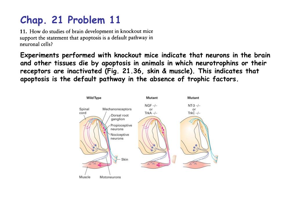
  1. Chap. 21 Problem 11 Experiments performed with knockout mice indicate that neurons in the brain and other tissues die by apoptosis in animals in which neurotrophins or their receptors are inactivated (Fig. 21.36, skin & muscle). This indicates that apoptosis is the default pathway in the absence of trophic factors.

  2. Chap. 21 Problem 12 Apoptosis is a regulated program that can be induced by withdrawal of trophic factors, which signal cells to stay alive, or by receipt of death signals like tumor necrosis factor, which trigger apoptosis. The structural changes that occur during apoptosis are morphologically distinct from changes that occur due to cell death via necrosis (Fig. 21.30). In necrosis, cells typically burst and release their contents outside. This damages surrounding cells and can lead to inflammation. In apoptosis, cells shrink, condense, and fragment without the release of cell contents. Cell fragments known as apoptotic bodies are later phagocytosed. Apoptosis proceeds via the same morphological events regardless of whether it is triggered by a death signal or by withdrawal of a trophic factor. This is because the same intracellular processes are triggered by the two types of signaling.

  3. Chap. 21 Problem 16 a) Bad is a pro-apoptotic factor that is activated in the absence of trophic factors (Fig. 21.38). Phosphorylation of Bad in the presence of a trophic factor inhibits its pro-apoptotic activity. Thus, a mutation that blocks Bad phosphorylation will cause cells to undergo apoptosis even in the presence of trophic factors. b & c) Cells that overexpress Bcl-2, an anti-apoptotic factor should not undergo apoptosis even in the absence of trophic factors. Likewise, Bax mutations that block its oligomerization should interfere with apoptosis. Both b & c types of mutations could contribute to cancerous transformation due to the fact that they block apoptosis even in the absence of trophic factors.