Download
1 / 22
220 likes | 408 Vues
The Female Reproductive System (Chapter 28). Lecture # 17 : The Female Reproductive System. Objectives. 1- To describe the structure and functions of the principal organs of the female reproductive system. 2- To describe the oogenesis.

E N D
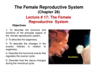
The Female Reproductive System (Chapter 28) Lecture # 17: The Female Reproductive System Objectives 1- To describe the structure and functions of the principal organs of the female reproductive system. 2- To describe the oogenesis. 3- To describe the changes in the ovarian follicles in relation to oogenesis. 4- Describe the hormonal events that regulates the ovarian cycle. Ovulation 5- Describe how the uterus changes during the menstrual cycle.
The Female Reproductive System Main Reproductive Organs or Gonads External Genitalia Accessory Glands and Organs Duct system Clitoris Uterine tubes Bartolini’s or greater vestibular glands Ovaries Labia minora Uterus Labia mayora Vagina Paraurethral glands Internal Genitalia External Genitalia
The Female Reproductive System Ovary Uterine tubes Uterus Vagina Paraurethral glands Clitoris Labia minora Bartolini’s or greater vestibular glands Labia mayora
The Female Reproductive System is more complex than that of the male because it serves more purposes: 1- It produces and delivers gametes. 2- It provides nutrition and safe harbor for fetal development. 3- It gives birth, and nourishes the infant. The Ovaries The egg develop in their own fluid-filled follicles They are the female gonads which produce egg cells (ova) and sex hormones. It is occupied by major arteries and veins. Medulla • It is where germ cells develop. • Follicle bursting and releasing the egg (ovulation). Cortex Tunica albuginea
The Uterine Tubes The Ovarian Ligaments Ovarian ligament Ampulla Isthmus It attaches the ovary to the uterus. Suspensory ligament • It attaches the ovary to the pelvic wall and contains ovarian artery, vein and nerves. Broad ligament Fimbriae Infundibulum Broad ligament: Functions: Mesosalpinx They receive and transport the egg to the uterus. Mesovarium It attaches the ovary to the broad ligament. It is the site where fertilization generally occurs. Mesometrium
Uterine tubes are lined by a simple columnar epithelium with ciliated cells and a smaller number of secretory cells. The cilia beat toward the uterus and, with the help of muscular contraction of the tube, convey the egg in that direction.
Once it has been released, the oocyte begins a slow journey to the uterus through the uterine tubes (fallopian tubes). Ovulation Implantation DAYS 7-10 While within the tube, the oocyte may encounter sperm and become fertilized prior to entering the uterus.
The Uterus It is a thick muscular chamber that opens into the roof of the vagina. Functions: 1- It harbors and provides a source of nutrition for the fetus. • 2- It expels the fetus at the end of its development. Fundus Perimetrium It is the external serosa layer. Myometrium • It constitutes most of the uterine wall, and is composed mainly of smooth muscle. Body • It produces labor contractions and expels the fetus. Endometrium It is a simple columnar epithelium, compound tubular glands, and a stroma populated with leukocytes, macrophages, and other cells. Internal os • Stratum functionalis: The superficial half, shed each menstrual period. Lateral fornix Cervix Cervical canal External os • Stratum basalis: The deep layer, stays behind and regenerates a new stratum functionalis with each menstrual cycle. • During pregnancy, the endometrium is the site of attachment of the embryo and forms the maternal part of the placenta from which the fetus is nourished. Vagina
The Vagina It is a distensible muscular tube (8-10 cm). Functions: 1- It is a passageway for menstrual flow. Posterior fornix Uterus 2- It receives the penis and semen. Anterior fornix 3- It is a passageway for delivery. Urinary bladder • The vaginal epithelium is simple cuboidal in childhood, but the estrogens of puberty transform it into a stratified squamous epithelium (vaginal metaplasia). Rectum Urethra Vagina Urethral orifice • Bacteria ferment glycogen rich cells producing acidic pH in vagina, which inhibits the grow of pathogen. Vaginal orifice Anus They keep the vulva moist, and provide lubrication during sexual excitement. Bartolini’s or greater vestibular gland Paraurethral gland
Mons pubis Prepuce Urethral opening Glans or clitoris Hymen (torn) Labia minora Vaginal entrance Labia majora External Genitalia Perineum
Lactiferous duct Lactiferous sinus Lobules Suspensory ligament Nipple Areola The Mammary Glands By the end of the six month of pregnancy the mammary glands are fully developed (prolactin hormone produced by the adenohypophysis). They produce milk to nourish the baby. Pectoralis major After birth Pectoral fat pad Lobes
Puberty and Menopause • Puberty begins at age 8-10 for most girls in US. Gonadotropin releasing hormone (GnRH) • It is triggered by rising levels of Gonadotropin releasing hormone (GnRH) produced by the hypothalamus. • GnRH stimulates the anterior lobe of pituitary to produce gonadotropins: • Follicle-stimulating hormone (FSH) • Follicle-stimulating hormone (FSH) • Luteinizing hormone (LH) Inhibin • Luteinizing hormone (LH) • FSH stimulates developing ovarian follicles and they begin to secrete estrogen, progesterone, and inhibin. Estrogens • Estrogens are feminizing hormones with widespread effects on the body. The principal estrogens are : Estradiol (the most abundant), estriol, and estrone. Progesterone
Feminizing Hormones Estradiol is the most abundant estrogen (feminizing hormones). Effects of the Estradiol: • 1- It stimulates vaginal metaplasia. • The vaginal epithelium changes from simple cuboidal to stratified squamous. • 2- It stimulates growth of ovaries and secondary sex organs. • 3- It stimulates growth hormone secretion. • 4- It produces an increase in height and widening of the pelvis. • 5- It is responsible for feminine physique because it stimulates the deposition of fat. • 6- It makes a girl’s skin thicker, but remains thinner, softer, and warmer than males of the corresponding age. Estradiol (estrogen)
Oogenesis and Sexual Cycle Sexual Cycle Reproductive Cycle The events that recur every month when pregnancy does not intervene. The sequence of events from fertilization to giving birth. • It consists of two interrelated cycles controlled by shifting patterns of hormone secretion: Follicular phase • Ovarian cycle Ovulation (The events in the ovaries) Luteal phase Proliferative phase • Menstrual cycle Secretory phase (The parallel events in the uterus) Premenstrual phase Menstrual phase
Events that Occur in the Ovaries 1- Oogenesis (production and development of oocytes). 2- Folliculogenesis (development of the follicles). 1- Oogenesis Like spermatogenesis, it produces haploid gametes by means of meiosis. Spermatogenesis 1- Males produce sperm continually. 2- Spermatogonia and spermatocytes are produce continually during fertile period of life. Oogenesis 1- It is a cyclic event that releases one egg per month. 2- Oogonia and primary oocyte are produce only before birth. • 3- Oogonia multiply until the fifth month reaching 5 to 6 million in number. Then go into a state of arrested development until shortly before birth. 4- Shortly before birth oogonia transform into primary oocyte. 5- Most of primary oocytes undergoes degeneration before the girl is born and during childhood. 6- By puberty, only about 400, 000 remains (even if a woman ovulates every 28 days from 14 to 50, she would ovulate 480 times.
1-Oogenesis Between the third and seventh month of fetal life: Oogonia Oogonia undergo mitosis and produce primary oocytes (diploid). Diploid OOGENESIS MITOSIS Primary oocytes (diploid) begin MEIOSIS I but it is stopped in prophase I. First Primary Second Secondary Secondary Diploid At birth: oocyte polar polar oocyte oocyte MEIOSIS I Primordial follicles Primary oocytes (diploid) in prophase I of MEIOSIS I. Before birth It stops in It stops in metaphase prophase At puberty: After puberty Primary follicles MEIOSIS II MEIOSIS I FSH triggers the start of the ovarian cycle. Completed Completed Secondary follicles Haploid MEIOSIS I is completed to form one secondary oocyte (haploid)and the first polar body. Ovulation Tertiary follicles body body MEIOSIS II Before ovulation During reproductive life: After ovulation Every month one secondary oocyte begins MEIOSIS II that is stopped in metaphase II. Haploid If fertilization Ovulation occurs, and if the secondary oocyte is fertilized, MEIOSIS II is completed to form the ovum and the second polar body. occurs
2- Folliculogenesis As the egg undergoes oogenesis, the follicle around it undergoes developments called folliculogenesis • They consist of a primary oocyte in early meiosis, surrounded by a single layer of squamous follicular cells. They are concentrated in the cortex of the ovary. • Primordial follicles They have larger oocytes and follicular cells that still form a single layer. • Primary follicles They have still larger oocytes and follicular cells are now stratified (granulosa cells). Zona pellucida: It is a layer of glycoprotein gel secreted by granulosa cells around the oocyte. • Secondary follicles • Theca folliculi: It is a fibrous husk of connective tissue around the granulosa cells. • Granulosa cells begin secreting follicular fluid, which accumulates in little pools in the follicular wall - this defines the tertiary follicles • Tertiary follicles • As they enlarge, the pools merge forming a single fluid-filled cavity, the antrum. • Normally only one follicle from each month’s cohort becomes a mature follicle destined to ovulate while the rest degenerate. • Mature (Graafian) follicles
Oogenesis and Sexual Cycle Ovarian cycle • Ovarian cycle Follicular phase Ovulation (The events in the ovaries) Luteal phase Menstrual cycle Menstrual phase • Menstrual cycle Proliferative phase Secretory phase (The parallel events in the uterus) Premenstrual phase
The Ovarian Cycle Follicle Stimulating Hormone (FSH) Gonadotropins It stimulates the development of the follicles and the secretion of estradiol. They are produced by the anterior pituitary. Luteinizing Hormone (LH) It stimulates ovulation. It is produced by the maturing follicles and stimulates secretion of LH and FSH by the anterior pituitary Estradiol LH Gonadotropin releasing hormone (GnRH) Progesterone It has a crucial role in preparing the uterus for the possibility of pregnancy. Estrogens Estradiol Pituitary FSH Follicular phase Luteal phase
Ovarian Cycle Follicular phase It is characterized by growing follicles. It is governed by follicle stimulating hormone (FSH) from the pituitary gland and estrogens from the ovarian follicles. Ovulation It is triggered by luteinizing hormone (LH) from the pituitary gland (released in response to estrogen secretion for the follicles). Luteal phase It is governed by the luteinizing hormone (LH) from the pituitary gland, and progesterone and estrogen produced by ovarian corpus luteum.
The Menstrual Cycle Secretory phase ( 15 to 26) Proliferative phase (6 to 14 day) The endometrium thickens still more in response to progesterone from corpus luteum. At the end of the menstruation, the endometrium consists only of the stratum basale. Stratum functionalis • The endometrium thickens as a result of secretion and fluid accumulation rather than mitosis. • As new cohort of follicles develop in the ovaries, they secrete more and more estrogen. • Progesterone stimulates endometrial glands to secrete glycogen. Glands grow wider, longer and more coiled. • Estrogen stimulates mitosis in the stratum basalis and the prolific regrowth of blood vessels regenerating the functionalis, By day 14 is 2 to 3 mm thick. Stratum basale • By the end of this phase, the endometrium is 5 to 6 mm thick- a soft, wet, nutritious bed available for embryonic development. Premenstrual phase ( 27 and 28 days) Menstrual phase (1 to 5 day) • The corpus luteum atrophies and progesterone levels fall sharply, triggering spasmodic contractions of spiral arteries. • When enough menstrual fluid accumulates in the uterus, it begins to be discharged by the vagina. • It causes endometrial ischemia and brings about tissue necrosis and menstrual cramps • Pools of blood accumulate in stratum functionalis and necrotic endometrium mixes with blood and serous fluid and forms the menstrual fluid. Spiral artery
Menstrual Cycle: Proliferative phase • Estrogen stimulates mitosis in the stratum basalis and the prolific regrowth of blood vessels regenerating the stratum functionalis. Secretory phase The endometrium thickens still more in response to progesterone from corpus luteum. Progesterone Premenstrual phase • Corpus luteum atrophies and progesterone levels fall sharply. This causes endometrial ischemia with tissue necrosis and menstrual cramps. Estradiol The first day of discharge is day 1 of the new cycle Menstrual phase Necrotic endometrium mixes with blood and serous fluid and produce the discharge of menstrual fluid from the vagina. Premenstrual phase Menstrual phase Proliferative phase Secretory phase


