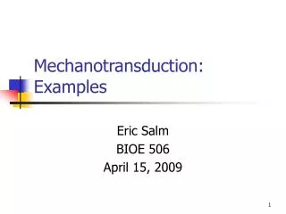
Cell Mechanotransduction
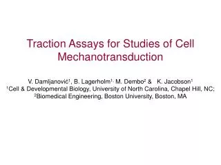
Traction Assays for Studies of Cell Mechanotransduction V. Damljanovi ć 1 , B. Lagerholm 1, M. Dembo 2 & K. Jacobson 1 1 Cell & Developmental Biology, University of North Carolina, Chapel Hill, NC; 2 Biomedical Engineering, Boston University, Boston, MA. Cell Mechanotransduction.

Cell Mechanotransduction
E N D
Presentation Transcript
Traction Assays for Studies of Cell MechanotransductionV. Damljanović1, B. Lagerholm1, M. Dembo2 & K. Jacobson11Cell & Developmental Biology, University of North Carolina, Chapel Hill, NC; 2Biomedical Engineering, Boston University, Boston, MA
Cell Mechanotransduction Signal Cytoskeleton Apply tractions Sense environment Motion Correlate w/ cell state Feedback
Cell Tractions Direction of migration “Propulsive” tractions “Frictional” tractions Elastic substrate Substrate tractions Adhesion molecules Tractions are determined from the deformation of substrate Polyacrylamide gel on 22 x 22 mm coverslip (modified protocol of Yu-li Wang)
Experimental Requirements(must match theoretical assumptions) • Gel must be flat, with free edges and bottom fixed on the coverslip • Gel thickness must be orders of magnitude greater than average displacement, but small enough for optics (70-100mm optimal) • Fluorescent markers must be small (we use 0.2mm) and only at the top (not really the case) • Must keep the focus always at the same set of beads (difficult) • Must be isolated from all vibrations—translation is tolerable, but not rotation
Swelling in fluid % acrylamide and BIS Media ionic content Conditions That Affect Gel Modulus
Deformed Cell applies tractions Stress-free Top view Assay Basics Displacement map Bead positions (fluorescence) Integration contour From Theory of Elasticity calculate Cell Tractions Cell shape (phase) (null image) – (deformed image) = (displacement map)
R2 R1 R2 R2 more affordable, easier to use and provides more consistent coating than previously used UV-activatable x-linker Sulfo-SANPAH Hydrazine hydrate (reducing agent) NH2 NH2 Activated polyacrylamide O C H2N — NH2 NH Hydrazine hydrate PA PA Polyacrylamide R1 N C Hydroxylysin in collagen R1 R1 NH R2 C C O PA NaIO4 OH H C Oxidized collagen Collagen-coated gel Conjugation of ECM Proteins to PA Gels
Correspondence Failures in Correlation-Based Optical Flow 1.4mm lower 0.6mm lower Gel top Good correspondence with null-image No correspondence with null-image Slight shift of focus plane results in loss of relevant displacement field Must always capture images of the same TOP bead layer
25mm 10mm PDMS stamp Micro-patterning With ECM Proteins • Excellent results, patterns from 5mm to few 100 mm • Instant transfer, despite of stamp slipping due to alignment by hand 30 sec Fluorescently labeled protein PA gel H-h activated Cells on 25mm stripes Cells on flat-printed area
19.4 mm 6.71 kPa Traction shear (mag. of traction gradient) Strain energy density (traction * displacement) Traction vectors Traction magnitude x 0.1 kPa x 10-7 J/m2 x 0.1 kPa/cm “Control” C3H Pax (-/-) MEF Paxillin and Mechanotransduction • Working on Pax (-/+) MEF & wild type MEF tractions control • Overexpress zyxin, vinculin or FAK, try to recover motility
Leading Edge Ruffles Both Push and Pull Ruffles are free (no FAs) and used for probing alternately push and pull on the substrate 2 min apart • One more proof of two distinct actin networks: • - Strip along the leading edge has no FAs, • can push and pull to probe • Inner part, behind leading edge has FAs • and always pulls
C3H (phase) in the moment of hesitation Green fluorescent collagen stripes Red fluorescent beads at the gel surface ?? Traction magnitude Traction vectors x 0.1 Pa 1030 kPa 21.7 mm 1-D Constrained MigrationWhat Controls Cell Direction and Polarity? Used m-patterned gel to: • Geometrically enforce cell polarity • & unidirectional migration • Simultaneously record tractions • and process of changing direction Future work: • Perturb leading edge (end of stripe, CALI, photoactivation) • Record protein activity
Hypothesis: HGF does not directly disrupt E-cadherin function. It increases integrin-mediated ECM adhesion The force of cells pulling apart breaks the junctions 10.4 mm HGF x 0.1 Pa 0 30 45 60 75 90 HGF and CadherinMechanism of MDCK spreading Collaboration with Martin Schwartz, UVa Time [min]