Download
1 / 52
520 likes | 644 Vues
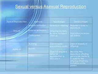
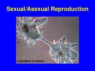
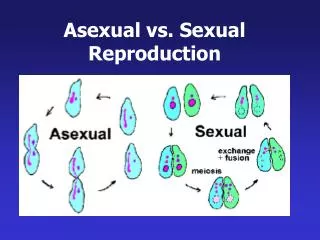
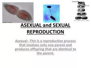
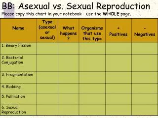
Fig. 30.1 Asexual reproduction in Euglena. 30.1 Asexual and Sexual Reproduction. In asexual reproduction , the offspring are genetically identical to one parent. The process begins with mitosis Protists typically divide by fission. Cnidarians typically divide by budding.

E N D
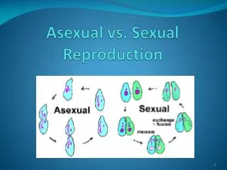
Fig. 30.1 Asexual reproduction in Euglena 30.1 Asexual and Sexual Reproduction In asexual reproduction, the offspring are genetically identical to one parent • The process begins with mitosis • Protists typically divide by fission • Cnidarians typically divide by budding
30.1 Asexual and Sexual Reproduction • In sexual reproduction, a new individual is formed by the union of two gametes (egg and sperm) • A zygote is formed • Develops by mitosis into a multicellular organism • Haploid gametes are produced in the gonads
Different Approaches to Sex • Parthenogenesis • Offspring are produced from unfertilized eggs • Common among arthropods • Some are exclusive • Others switch! • Common also in some lizard species
Hamlet bass Fig. 30.2 Bluehead wrasse • Hermaphroditism • One individual has both testes and ovaries • Tapeworms and earthworms Switch sexual roles! • Hermaphroditismmay be sequential • Individuals change sex Switch sexes! • Protogyny • From female to male • Protandry • From male to female
Sex Determination • In some reptiles, sex is determined by environmental changes • In mammals, it is determined early in embryonic development • Embryonic gonads are indifferent • Y chromosome converts them to testes • Responsible gene is SRY • Sex-determining region of the Y chromosome
30.2 Evolution of ReproductionAmong the Vertebrates • Vertebrate sexual reproduction evolved in the ocean before vertebrates colonized land • Most marine bony fish use external fertilization • Male and female gametes are released into the water where fertilization occurs • Most other vertebrates use internal fertilization • Male gametes are introduced into the female reproductive tract
There are three strategies for internal fertilization • 1. Oviparity • Fertilized eggs are deposited outside mother’s body to complete their development • 2. Ovoviviparity • Fertilized eggs are retained within the mother to complete their development • Young obtain nourishment from egg yolk • 3. Viviparity • Fertilized eggs are retained within the mother to complete their development • Young obtain nourishment from mother’s blood
Fish and Amphibians • The eggs of most bony fish are fertilized externally • Eggs contain little yolk • Young fish must seek its food from the water surrounding it • Thousands of eggs are fertilized, but only a few of resulting individuals reach maturity • Fertilization in most cartilaginous fish is internal • Development of young is viviparous
Fig. 30.6 Poison arrow frog South American marsupial frog Fig. 30.7 Surinam frog Darwin’s frog • Fertilization is external in most amphibians • Eggs of most species develop in water • With some interesting exceptions Male! Female • Development is divided into embryonic, larval and adult stages Female Male!
Fig. 30.8 How turtles do it Reptiles and Birds • Most reptiles are oviparous, laying amniotic eggs • Other species are ovoviviparous or viviparous • Most male reptiles use a penis to inject sperm into females • This process is called copulation
Fig. 30.9 • All birds are oviparous, laying amniotic eggs • As egg passes along oviduct, glands secrete albumin proteins and the hard calcareous shell • Birds are homeotherms • Incubate eggs to keep them warm
Mammals • Some mammals are seasonal breeders • Others have reproductive cycles • Periodic release of a mature ovum (ovulation) • Most female mammals have estrous cycles • Females sexually receptive to males only around time of ovulation (estrus) • Apes and humans have menstrual cycles • Females bleed when shedding inner lining of the uterus • Can copulate at any time in their cycle • Cats and rabbits are induced ovulators • Ovulation only after copulation due to LH secretion
Three types of mammals • Monotremes are oviparous • Lay eggs • Young hatchlings obtain milk by licking mammary glands (they lack nipples) • Marsupials are viviparous • Give birth to incompletely developed fetuses • Complete development in mother’s pouch • Obtain food from nipples in mammary glands • Placentals are viviparous • Retain young in uterus for long periods of development • Fetuses are nourished by the placenta
Duck-billed platypus Kangaroo Deer Fig. 30.10 Reproduction in mammals
Fig. 30.11 The male reproductive organs 30.3 Males
Fig. 30.13 Human sperm cell • The testis producessperm and testosterone • Enclosed in a hanging sac called the scrotum • Sperm need cooler temperature to develop • Spermatogenesis occurs in the seminiferous tubules • Sperm are then transferred to the epididymis for storage and maturation • From there to the vas deferens • To the urethra which empties through the penis Contains 23 chromosomes
Fig. 30.14 • The penis contains long cylinders of spongy tissue • These get filled with blood causing an erection Physical stimulation is required for ejaculation • 2-5 milliliters of semen are ejected • This volume contains several hundred million sperm • Plus secretions from the prostate and other glands
Fig. 30.15 The female reproductive organs 30.4 Females
At birth, a female’s ovaries contains all the oocytes she will ever produce • ~ 2 million oocytes are arrested in prophase I of the first meiotic division • At puberty, the release of FSH causes the resumption of meiosis I in a few oocytes • However, only one becomes dominant and is ovulated • Mature egg cells are called ova (singular, ovum) • This cycle is repeated about every 28 days
Fertilization of the egg occurs high in the Fallopian tubes (also called uterine tubes or oviducts) • The fertilized egg is now called a zygote • It is transported to the uterus • A muscular pear-shaped organ about the size of a fist • It narrows to a muscular ring called the cervix • Leads to the vagina
Fig. 30.17 A comparison of mammalian uteruses Primates Cats, dogs and cows Rats, mice and rabbits • Marsupials, such as opossums, have two unconnected horns, two cervices and two vaginas • Male marsupials have a forked penis!
The fertilized egg is pushed down the oviducts by the rhythmic contraction of its smooth muscles • The journey takes 5-7 days • The uterus is lined with a stratified epithelial membrane called the endometrium • The zygote attaches to this layer and begins embryonic development! • If the egg is not fertilized, the surface layer of the endometrium is shed during menstruation • The underlying layer generates a new surface layer during the next cycle
30.5 Hormones Coordinatethe Reproductive Cycle • The female reproductive cycle is composed of two distinct phases • Follicular phase • Egg reaches maturation and is ovulated • Luteal phase • Body continues to prepare for pregnancy • A family of hormones coordinates these two phases
Follicular Phase • Development of the egg within the ovary • The oocyte and its surrounding mass of tissue is called the follicle • FSH secretion triggers the maturation of several follicles and resumption of meiosis in their oocytes • But only one achieves full maturity • FSH also causes the ovary to secrete estrogen • Negative feedback by estrogen, causes the hypothalamus to stop the pituitary’s FSH output
Luteal Phase • The body is prepared for fertilization • Hypothalamus causes the anterior pituitary to begins secreting luteinizing hormone (LH) • LH inhibits further estrogen production • It also causes the wall of the follicles to burst • Oocyte is ovulated into oviducts • LH directs the repair of the ruptured follicle, which becomes the corpus luteum
Pituitary gland Levels of gonadotropic hormones in blood LH FSH FSH 0 7 14 21 28 days Ovarian cycle Developing follicles Ovulation Corpus luteum Luteal phase Hormone blood levels Estradiol Progesterone 0 7 14 21 28 days Endometrial changes during menstrual cycle Menstrual phase Proliferative phase Ovulation Secretory phase 14 21 28 days 0 7 Fig. 30.18 The human menstrual cycle
Luteal Phase • The corpus luteum begins to secrete the hormone progesterone • Progesterone inhibits FSH • It also thickens the endometrium preparing for fertilization • If fertilization does not occur, progesterone production stops and the luteal phase ends • Thickened endometrial layer sloughs off • This causes the bleeding associated with menstruation
Pituitary gland Levels of gonadotropic hormones in blood LH FSH FSH 0 7 14 21 28 days Ovarian cycle Luteal regression Developing follicles Ovulation Corpus luteum Luteal phase Hormone blood levels Estradiol Progesterone 0 7 14 21 28 days Endometrial changes during menstrual cycle Menstrual phase Proliferative phase Ovulation Secretory phase Menstrual phase 14 21 28 days 0 7 Fig. 30.18 The human menstrual cycle
Luteal Phase • If fertilization does occur, the corpus luteum is maintained by human chorionic gonadotropin (hCG) • hCG is a hormone produced by the embryo • It is tested for in all pregnancy tests • Two other hormones are of importance • Prolactin • Stimulates milk production • Oxytocin • Initiates milk release • Induces labor
30.6 Embryonic Development • The vertebrate embryo develops in three stages • Cleavage • A hollow ball of cell forms • Gastrulation • Cells move to the interior, forming the primary tissues • Neurulation • The organs of the body form
Cleavage: Setting the Stage for Development • During cleavage, zygote rapidly divides into larger and larger numbers of smaller and smaller cells • A morula forms • A tightly packed mass of about 32 blastomeres • Further division results in a hollow ball of 500-2,000 cells called the blastocyst • Contains a fluid-filled cavity, the blastocoel • Inner cell mass Forms the embryo • Trophoblast Becomes the placenta
Cleavage: Setting the Stage for Development • Embryo reaches the uterus on day 6 • It penetrates the endometrial lining • Initiates membrane formation • Amnion • Encloses embryo • Chorion • Forms from the trophoblast • Interacts with uterine tissue to form the placenta
Gastrulation: Onset of Developmental Change • Certain groups of cells move inwards from the inner cell mass at about 10-11 days after fertilization • This process of gastrulation results in the three primary germ layers • Endoderm • Ectoderm • Mesoderm
Neurulation: Determination of Body Architecture • In the third week, the three primary germ layers begin development into body tissues and organs • First, the notochord develops from the mesoderm • The neural tube develops from the ectoderm • The gut develops from the endoderm • On either side of the notochord blocks of tissue form • These somites give rise to muscles, vertebrae and connective tissues developing notochord • By the end of the third week, the embryo is about 2 mm (< 0.1 inches) long
Fig. 30.20a 30.7 Fetal Development Fourth week • Formation of body organs, or organogenesis • Critical time in development • Alcohol use may cause fetal alcohol syndrome • Embryo reaches about 5 mm
Second month Fig. 30.20b 30.7 Fetal Development • Great changes in morphology occur • Limbs assume adult shape • Major internal organs are evident • Embryo reaches about 25 mm
Third month Fig. 30.20c 30.7 Fetal Development • Development is essentially complete except for lungs and brain • Developing human is now called a fetus • It carries out primitive reflexes like sucking
Second trimester Fig. 30.20d 30.7 Fetal Development • A time of growth • Bone formation occurs • Hair and body are covered with fine hair called lanugo • By the end of the 6th month, the fetus is 30 cm (1 foot) long
Fig. 30.21 30.7 Fetal Development Third trimester • Pace of growth accelerates • Weight of fetus more than doubles • Nutrients provided by mother’s blood via the placenta • Most major nerve tracts are formed in the brain
Fig. 30.22 Postnatal development • Babies typically double birth weight within a few months • Different body parts grow at different rates • Allometric growth • Nerve cells produced at an average rate of > 250,000 per minute • At 6 months, neuron production ceases permanently
30.8 Contraception and Sexually Transmitted Diseases • Contraception, or birth control, is the prevention of pregnancy • Several different methods are available • These differ in their effectiveness and acceptability to different couples
Abstinence • Simplest and most reliable way • Natural family planning, or the rhythm method • Prevention of egg maturation • Birth-control pills • Estrogen and progesterone • Shut down production of the pituitary hormones FSH and LH • Birth-control injections • Birth-control patches
Prevention of embryo implantation • Intrauterine devices (IUDs) • RU486 (“morning after pill”) • Sperm blockage • Condoms • Diaphragms • Sperm destruction • Spermicidal jellies • Foams • Sterilization • Vasectomy in males • Tubal ligation in females