Download
1 / 22
230 likes | 601 Vues
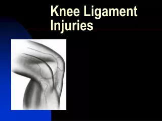
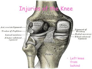
Radiographic Imaging in Acute Knee Injuries. Nick Sparler MS4. Topics. Basic Anatomy To Image or Not? Bony injury Ligamentous injury. Anatomy. To Image or Not? Fracture?. Ottawa knee rules Age 55 years or older Tenderness at head of fibula Isolated tenderness of patella

E N D
Radiographic Imaging in Acute Knee Injuries Nick Sparler MS4
Topics • Basic Anatomy • To Image or Not? • Bony injury • Ligamentous injury
To Image or Not?Fracture? Ottawa knee rules • Age 55 years or older • Tenderness at head of fibula • Isolated tenderness of patella • Inability to flex knee to 90 degrees • Inability to walk four weight-bearing steps immediately after the injury and in the emergency department Pittsburgh decision rules • Blunt trauma or a fall as mechanism of injury plus either of the following: • Age younger than 12 years or older than 50 years • Inability to walk four weight-bearing steps in the emergency department * In a recent prospective study, the Pittsburgh decision rules were 99 percent sensitive and 60 percent specific for the diagnosis of knee fractures. The Ottawa knee rules were 97 percent sensitive and 27 percent specific for knee fractures
To Image or Not?Ligament? • History and Physical exam • Mechanism or injury • Lachman test, Anterior Drawer test, Gravity test etc • MRI is the preferred diagnostic modality
Bony injuries • AP, Lateral and Oblique views should be obtained • Lateral tibial plateau (TP) fractures are most common • Patellar fractures due to trauma or forced contraction of quadriceps tendon • Beware of Bi-Partite Patella • Segond- avulsions of the LCL at Lat TP • Think ACL or Meniscus
ACL Injurywith Bony Fracture1. Hemarthrosis ACL 2. Avulsion 3. Segond Fracture
Ligamentous Injury • ACL most common • PCL- “dashboard injury”, hyperextension associated with ACL injury • MCL/LCL • Meniscus
Grading • - Grade I (microscopic tears) ligamentous tears demonstrate an intact ligament of normal thickness surrounded to a variable degree by intermediate T1-weighted and high T2-weighted signals indicative of surrounding edema. The ligament remains closely applied to the underlying cortical bone. • - Grade II (partial tears) tears demonstrate thickening and/or partial disruption of the fibers of the MCL with an increased amount of surrounding intermediate T1-weighted and high T2-weighted signals indicative of increased edema and concomitant hemorrhage. • - Grade III (complete tear) tears demonstrate complete disruption of the ligament with corresponding surrounding hemorrhage and edema.
ACL Injury • Confirm in the coronal and axial planes
Resources • Grainger and Allison’s Diagnostic Radiology • http://www.wikiradiography.com/page/Radiographic+Anatomy • www.sportsdoc.umn.edu/.../normal%20PCL%20mri.jpg • http://www.aafp.org/afp/991201ap/2599.html • www.mskcases.com