Extraocular Muscles (EOM) & Eye movement
560 likes | 1.15k Vues
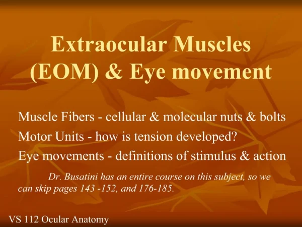
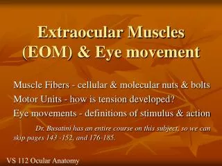
Extraocular Muscles (EOM) & Eye movement. Muscle Fibers - cellular & molecular nuts & bolts Motor Units - how is tension developed? Eye movements - definitions of stimulus & action Dr. Busatini has an entire course on this subject, so we can skip pages 143 -152, and 176-185.

Extraocular Muscles (EOM) & Eye movement
E N D
Presentation Transcript
Extraocular Muscles (EOM) & Eye movement Muscle Fibers - cellular & molecular nuts & bolts Motor Units - how is tension developed? Eye movements - definitions of stimulus & action Dr. Busatini has an entire course on this subject, so we can skip pages 143 -152, and 176-185. VS 112 Ocular Anatomy
I. Just what sort of movements do your eyes make? • Those which are you aware of, those that are voluntary (or at least the initiation of them is voluntary). • Reflex eye movements - automatic adjustment of the eye position to stabilize an image on the eye.
Eye movements • Smooth tracking (pursuit) • Combined head and eye tracking • Saccades – quick jumps, saccadic eye movements (< 50 msec) (up to 20°) voluntary initiation • Mini saccades (see fixation). • Fixation – holding a steady gaze (which we never actually do – see figure 4.1).
Reflex eye movements. The stimulus can be head/body movement • i) VOR vestibular ocular reflexes – if you move your head the eyes can maintain fixation upon the target. • ii) Optokinetic reflex : stimulus = rapid motion of the world, such as motion of a stream, or motion of world outside of the side car window. • Nystagmus (alternating slow & quick phases of movement)
Reflex eye movements.The stimulus can be movement, motion of an object of interest. • iii) Smooth pursuit - one object is targeted and remains clear on the retina the rest of the visual world is in motion – the standard stimulus for the optokinetic reflex,(only seen in foveate animals) • iv) Fixation - holding a steady gaze on a target of interest. NOT just a special form of pursuit.
Fixation – holding a steady gaze • The eyes are in constant motion • Slow drift • Minisaccades • Tremor (high frequency jitter in position) • aka micronystagmus. • In fact, if you could perfectly stabilize an image on the retina it would fade and disappear. Some retinal motion is necessary, that is some movement of the image across the retina must be present or the image fades.
Eye Movements: • See figure 4.1 over head
Motion of an Eye • To describe eye motions we need a set of defined axes (Fick’s Axes - draw on board) • X axis : nasal -> temporal • Y axis: anterior -> posterior • Z axis: superior -> inferior • These axes intersect at the center of rotation - a fixed point, defined as 13.5 mm behind cornea.
Ductions (single eye movements) • Rotation about the Z axis (Z axis runs vertically superior to inferior) • Medial Rotation - adduction toward midline • Lateral Rotation - abduction away from midline • Rotation about the X axis (X axis runs horizontally, from nasal to temporal) • Upward, elevation (supraduction) • Downward, depression (infraduction)
Torsion - cyclorotations • Rotation about the Y axis (Y axis runs horizontally, from anterior to posterior) • These are described with respect to a point at 12 o‘clock on the superior limbus • Intorsion (incyclorotation) rotation nasally • Extorsion (excyclorotation) rotation of the 12 o’clock position temporally. • Counteracting head tilt (up to 7-9°)
Version & Vergences • Some eye movements are paired, that is both eyes do the same thing. . . . Versions • Sometimes eyes move in the opposite directions simultaneously. . . Vergences
Vergences • Disjunctive eye movements (opposite left- right movments). Non-yolked motion • Convergence (simultaneous movement nasally) • Divergence (simultaneous temporal movement) • Encyclovergence (intorsion) • Excyclovergence (extorsion)
Versions (conjugate eye movement) • Dextroversion - rightward gaze (demo) • Levoversion - leftward gaze • Supraversion - elevation • Infraversion - depression • Also up and right, up and left • Down and right, down and left • ALL BEHAVIOR IS THAT OF YOLKED EYES
Extraocular Muscles • 4 rectus muscles - origin is in the common tendous ring (annulus of Zinn) • Oval ring of connective tissue • Continuous with periorbita • Anterior to optic foramen • Medial and lateral rectus attached to both the upper and lower tendon limbs • The muscles traveling from the this tendon ring to the insertions create muscle cone.
Spiral of Tillaux • The rectus muscle pass through tenon’s capsule and insert into the sclera. • The muscles insert at different distances from the cornea. • The insertion pattern is a spiral with the medial rectus closest to the cornea (5.5 mm) and the superior rectus the furthest away from the cornea (7.4 mm).
Medial Rectus • Originates on both the upper and lower limb of the common tendous ring and the optic nerve sheath. • Inserts along a vertical line 5.5 mm from the cornea. The horizontal plane of eye bisects the insertion. • Fascial expansion from muscle sheath forms the medial check ligament and attach to medial wall of orbit.
Medial Rectus cont. • Innervation is via cranial nerve III, the oculomotor nerve, and the specific branch runs along the inside of the muscle cone, on the lateral surface. • The superior oblique, ophthalmic artery and nasociliary nerve all lie above the medial rectus.
7.4 mm 6.9 mm 6.7 mm Spiral of Tillaux 5.5 mm
Lateral rectus • Originates on both the upper and lower limb of the common tendous ring. . .AND a process of the greater wing of the sphenoid bone. • Inserts parallel to medial rectus 6.9 mm from the cornea. (Tendon 9.2 mm wide, 8.8 long). • Fascial expansion from muscle sheath forms the lateral check ligament and attach to lateral wall of orbit at Whitnalls tubercle.
Lateral Rectus cont. • Innervated by the abducens nerve, Cranial n VI which enters the muscle on the medial surface. • The lacrimal artery and nerve run along the superior border. • The abducens n., ophthalmic artery and ciliary ganglion lie medial to the lateral rectus and between it and the optic nerve.
Superior Rectus • Originate on superior limb of the tendonous ring, and optic nerve sheath. • Muscle passes forward underneath the levator, but the two sheaths are connected resulting in coordinated movements. • Insertion 7.4 mm from limbus, and obliquely. • The angle from the origin to the insertion is 23° beyond the sagital axis. (see overhead)
Superior Rectus cont. • Frontal nerve runs above the s. rectus & levat. • The nasociliary nerve and ophthalmic artery run below. • The tendon for insertion of the superior oblique muscle runs below the anterior part of the superior rectus. • Innervationis via superior division of CN III, from the inferior surface; additional branches make their way to the levator.
Action of Superior Rectus • Primary action is elevation . . But since the insertion on the globe is lateral as well as superior, contraction will produce rotation about the vertical axis toward midline • Thus secondary action is adduction • Finally, because the insertion is oblique, contraction produces torsion nasally Intorsion. • (overhead figure 10-13A)
Superior view of Sup. Rectus 23° Because the muscle runs at an angle to the Fick’s axes, contraction is not confined to one axis
Inferior rectus • Originates on lower limb of common tendonous ring. • Inserts 6.7 mm from limbus, insertion is an arc • It is parallel to superior rectus, making a 23° angle beyond the sagittal axis. • Innervated by inferior division of CN III which runs above it (within the muscle cone). • Below is the floor of the orbit and inf. oblique
Inferior Rectus cont. • Fascial attachments below attached to inferior lid coordinate depression and lid opening. • Fascia below Inf. Rectus and Inf. Oblique contribute to the suspensory ligament of lockwood. • Primary Action downward gaze depression • 2° Adduction, as is the case for sup. Rectus • Also extorsion due to oblique arc of insertion.
Superior Oblique • Anatomical origin is on the lesser wing of the sphenoid bone. The physiological origin is the trochlea, a cartilagenous “U” on the superior medial wall of the orbit. • Longest thinnest EOM, the muscle ends before the trochlea, tendon is 2.5 cm, smooth movement through trochlea. • Innervation by CN IV, the trochlear nerve posterior in the orbit.
Action of Sup. Oblique • Primary action is intorsion _ rotation of 12 o’clock position toward midline. • Because the insertion of the oblique muscle is in the lateral, posterior quadrant the secondary actions are • Rotating the back half of the globe from lateral to medial (the anterior pole will move away) ABDUCTION • Also depression (posterior superior quadrant of the globe being pulled upward).
Inferior Oblique • Originates on the maxillary bone inferior to the nasolacrimal fossa. The ONLY EOM originating in the anterior orbit. • Inserts on the posterior lateral aspect of globe mostly inferior, below the ant.-post. horizontal plane. • Innervation from inferior division of CN III inserts on the upper surface (within muscle cone.)
Action of Inf. Oblique • Primary is extorsion • 2° is due to posterior, lateral, inferior insertion being pulled around, underneath globe and toward the anterior inferior insertion medially. • Rotation about the Z axis will be nasal to temporal (abduction). • Rotation about the X axis will be elevation (see overhead figure 10.14)
And from here it’s complicated • There is a balance of tension in the pairs of muscle to start with. . . . • Actions of the muscle can and do depend on the starting position. • For example elevation of the eye in the straight-ahead position and lateral position is accomplished by the sup. rectus. • But when the eye is medial rotated elevation is accomplished by the inferior oblique muscle action. (see fig 10-17 over head).
Thick & thin filaments Elements within, between the Z-lines make up a contractile unit
Myosin head binds to actin filament. The ratchet motion moves the two filament about 12 nm with respect to each other. It takes only 5 ms Because of the large number of z-line segments or contractile units along a fiber, a fast motion is attained. Ratchet Model
Thick and thin filament specializations with EOM Thick Thin Each motor fiber is innervated by only one nerve
Extraocular muscles are special A motor nerve can and does contact more than one fiber usually 100’s The motor units are small, with only from 5 to 18 muscle fibers contact by each motor nerve
THICK FIBERS Striated muscle Singly innervated 1 nerve, 1 branch Motor end plate Terminaison en plaque Includes both fast and slow twitch fibers All or none contraction THIN FIBERS Striated musle Multiply innervated Many branches 1 nerve En grappe end plate Terminaisons en grappe Thought to be slow sustained (tonic) Graded contractions EOM’s are special
Thick and thin filament specializations with EOM Each motor fiber is innervated by only one nerve
A single nerve impulse • Generates a muscle AP and contraction - tension. • Multiple nerve AP’s • Tensions sum over time • Repetitive firing results in a sustained contraction. • Tetanus (unresolved individual twitches).
3 Basic types of motor units Slow Fast-fatigue resistant Fast- fatigable
Cat gastroc muscle • ATPase activity at neurtral pH • ATPase activity at acidic pH • NADH dehydrogenase stain
Recruitment within the motor nerve • Groups of cell bodies innervating the same muscle make up a MOTOR NUCLEUS • . . . .and give rise to a motor nerve. • Nerves within a motor nerve are activated in a characteristic sequence - the size principle. • Smaller fibers, fire first, and larger later • There is a characteristic pattern of recruitment, tension is added and removed in a repeated pattern.
Muscle Spindle & Golgi Tendon Organs • Specialized sensory organs within the muscle provide feed back to the brain. • How much tension is in the muscle? • Is there any stretch imposed? What is muscle length? • EOM’s do not have the typical stretch reflex