Download

1 / 62
680 likes | 1.3k Vues
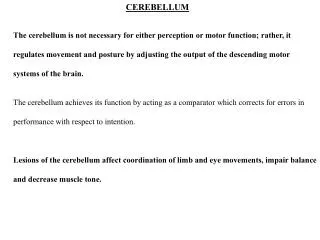
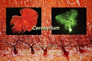
THE CEREBELLUM CEREBELLUM. Institute of Anatomy, 2nd Medical Faculty R. Druga. Cerebellar gyri = folia Cortex, subcortical white matter, nuclei. Gyri = folia, Cortex- podkorová bílá hmota – mozečková jádra. Excessive folding of the cerebellar surface (cortex). Little brain

E N D
THE CEREBELLUMCEREBELLUM Institute of Anatomy, 2nd Medical Faculty R. Druga
Cerebellar gyri = folia Cortex, subcortical white matter, nuclei Gyri = folia, Cortex- podkorová bílá hmota – mozečková jádra Excessive folding of the cerebellar surface (cortex)
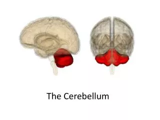
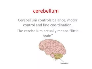
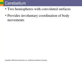
Little brain Located in the posterior cranial fossa Connected with the brain stem- by peduncles (inferior, middle, superior) Is covered by the cerebellar cortex (3 layers). Cortex is extensively folded (folia-oriented mediolaterally) In the white matter are the cerebellar nuclei The cerebellum – relations and structure Fossa cranii posterior Pedunculi cerebellares(inferior, medius, superior) Cortex – 3 vrstvy V bílé hmotě mozečková jádra
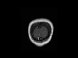
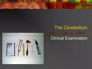
MR examination MR vyšetření Fossa cranii posterior Posterior cranial fossa
Arterial supply Arteriae cerebellares PICA AICA
Weigert staining
Marchi Staining Kluver – Barrera Marchiho metoda Kluver - Barrera
Unfolded surface of the cerebellum Dvourozněrná rekonstrukce povrchu (kůry) mozečku
Dvourozměrná Rekonstrukce lalůčků mozečku Dvourozměrní rekonstrukce lalůčků mozečku
Cerebellar lobes and lobuli Lobus - 3 Lobulus - 10 Vermis Paravermální oblast (zona) Laterální část hemisféry Mozečkové Laloky (3) a Lalůčky (10)
Mozečková jádra Neurony glutamátergní (excitační), vysoká spontánní aktivita CEREBELLAR NUCLEI, neurons glutamatergic, excitatory, high spontaneous activity
Mozečková kůra – 3 vrstvy Structure of the cerebellar cortex -3 layers:I.molecular layer- inhibitory interneurons I.Molekulární vrstva – inhibiční interneurony II. Purkyně cell layer – inhibitory projecting neurons II. Vrstva Purkyňových buněk – inhibiční, projekční neuronyIII. granular layer – prevail excitatory neurons III. Vrstva granulárních buněk – převaha excitačních neuronů
Šplhavá vlákna Olivocerebel. Projekce (zkřížená) Climbing Fibers Olivo-cerebellar projections (crossed)
Basket cells, inhibitory interneurons, GABAergic Košíčkové buňky, inhibiční interneurony
Mossy fibers – Granule cells- Paralel fibers –Purkyně cells Climbing fiber – Purkyně cell Šplhavá Vlákna – Purkyňovy b. Mechová Vlákna- granulární b. Paralelní vl.
Tracts Vestibulocerebellar (from the labyrinth and vestibular nuclei) Spinocerebellar ant., post., rostral, cuneocerebellar Olivocerebellar Reticulocerebellar Nucleocerebellar Pontocerebelar !! (cortico-ponto-cerebellar) Corticonuclear (from the cerebellar cortex to the nuclei) Vermis – nc. fastigii Paravermal zone – nc. embol. Nc. glob. Lateral hemisphere – nc. dentatus From cerebellar nuclei to the brain stem and to the thalamus Aferentní a eferentní spoje kůry mozečku Afferent and efferent connections
Spinocerebellar Pathways Posterior T1 – L2, uncrossed ICP, proprioceptors Anterior L3 – L5 Crossed, SCP, cutaneous signals Rostral C4 – C8 Uncrossed, ICP Cutaneous signals Cuneocerebellar C2 – T4 uncrossed Lateral cuneate nc., propriocept. ICP
Origin of the spinocerebellar pathways b a Dorsal (posterior) spinocerebellar projection- uncrossed Ventral (anterior) spinocerebellar projection - crossed
Funkční MR, flexe a extense ruky (červený, oranžový signál), nohy (modrý signál) FMI – increased blood flow during flexion and extension of ipsilateral hand (red, orange) and foot (blue) Zvýšení průtoku krve ve spinálním mozečku (lobus anterior)
Afferent projections Aferentní spoje
Cortico- pontine pathway, 17 millions fibers Neocortex – ipsilat. pontine ncc. - pontocerebellar pathway – contralateral cerebellar cortex (mossy fibers) Kortiko-pontinní dráha 17 milionů vláken Neocortex – ipsilaterální pontinní jádra Pontocerebellární dráha – Kontralaterální mozečková kůra (mechová vlákna)
Cortico-ponto-cerebellar pathway Cortico – ponto – cerebellar pathway
Climbing fibers Šplhavá vlákna Olivocerebellar projection
J. Eccles 1967 Nobel Prize in Physiology and medicine 1963
AFFERENTS TOTHE CEREBELLAR CORTEX I. • Climbing fibers – inferior olive (each P.cell receives only 1 c.f., many synapses with P.c.), excitatory (glutamate), firing frequency of the c.f. is very low (1 impulse/sec), c.f. elicit burst of action potentials in the P.c. • C.f. inform about errors in the execution of movements – error indicators !!
AFFERENTS TO THE CEREBELLAR CORTEX II • Mossy fibers - spinal cord, RF, pontine nuclei, ncc. cranial nerves. • End in the granular layer and each of which contacts large number of granular neurons. Granular cell axon contacts large number of P. c. via paralel fibers. • Mossy fibers are excitatory (glutamate). • Each mossy fiber influences many P.c. but the excitatory effect is weak. Many mossy fibers must be active together to provide sufficient excitation to fire a P.c. • Mossy fibers provide precisely graded information about movements, skin stimulations, joint position and about motor comands issued from the cerebral cortex.
Efferent connections of the cerebellar cortexCerebellar cortex – cerebellar nucleivermis – nc. fastigii, vestibular ncc. pavermal zone – emboliformis, globose ncc. lateral hemisphere – dentate nc.
Efferent connections of the cerebellar nuclei • Fastigial nucleus – vestibular nuclei, reticular formation • Emboliformis + globosusnucleus -.reticular formation, nc. ruber, thalamus • Nucleus dentatus – nc. ruber, contralateral thalamus (ventrolateral nucleus, intralaminar thalamic nuclei, ventral anterior nc., • Ventrolateral nucleus – primary motor cortex (area 4)
Thalamus Nc. Ventralis Lateralis Nc. VL – Motor cortex
Cortico-ponto-cerebello-cortical circuit (Transcerebellar Circuit)
Mozečkové syndromy • Vestibulární mozeček a vermis – poruchy rovnováhy, stoje a chůze, chůze o široké bazi, nystagmus • Spinální mozeček – kontroluje axiální svalstvo a proximální svaly končetin. Při poškození zvýšení tonu extensorů. • Pontinní mozeček (hemisféry) - přestřelování pohybů (hypermetrie, prst – lalůček, prst- špička nosu). Adiadochokinéza, třes(méně než 5 Hz- zhoršuje se na konci zacíleného pohybu)), poruchy řeči a výslovnosti (dysartrie, skandovaná řeč), poruchy plánování, uvažování (kognitivní poruchy).