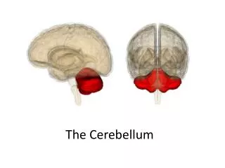
The Cerebellum
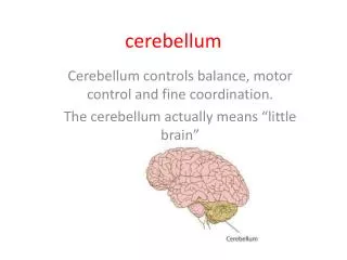
The Cerebellum. 陽明大學醫學 院 神經學系. 陳昌明 醫師. Position. Lies above and behind the medullar and pons and occupies posterior cranial fossa. Cerebellum. Cerebellum External Configurations. L ocated in posterior cranial fossa T entorium cerebelli (cerebrum), 4 th ventricle (brain stem)

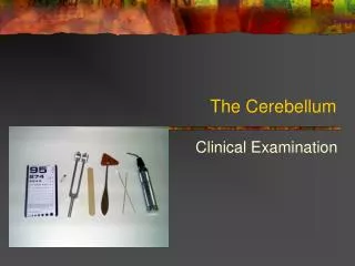
The Cerebellum
E N D
Presentation Transcript
The Cerebellum 陽明大學醫學院 神經學系 陳昌明 醫師
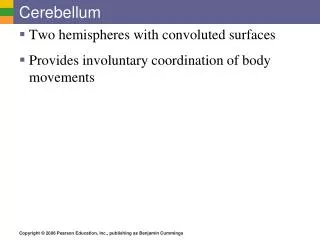
Position • Lies above and behind the medullar and pons and occupies posterior cranial fossa Cerebellum
Cerebellum External Configurations • Located in posterior cranial fossa • Tentorium cerebelli (cerebrum), 4th • ventricle (brain stem) • Communicate with other structure via • superior, middle, and inferior cerebellar peduncle
External features Consists of twocerebellar hemisphere united in the midline by the vermis
External features Three peduncles • Inferior cerebellar peduncle 下小腦脚 -connect with medulla and with spinal cord, contain both afferent and efferent fibers • Middle cerebellar peduncle 中小腦脚-connect with pons, contain afferent fibers • Superior cerebellar peduncle 上小腦脚-connect with midbrain, contain mostly efferent fibers
Cerebellum • Longitudinal division • Vermis, Paravermal Region, Cerebellar Hemisphere • Transverse division • Anterior Lobe • Posterior Lobe • Flocculonodular Lobe
Lobes Anterior lobe corpus of cerebellar Primary fissure Posterior lobe Flocculonodular lobe Posterolateral fissure
Lobes • Two deep fissures • Primary fissure • Posterosuperior fissure • Three lobs • Flocculonodular lobe 絨球小結葉flocculus and nodule • Anterior lobe • Posterior lobe Corpus of cerebellar
External features Superior surface • Tonsil of cerebellum小腦扁桃体two elevated masses on inferior surface of hemispheral portion just nearby foramen magnum Tonsil View from below
Functional divisions of cerebellar cortex Cbm unfold
External features Cerebellum & Brainstem, Inferior Surface, Anterior View
Cerebellum External Configurations Subdivision of Flocculonodular Lobe Nodulus Flocculus Subdivision of Anterior Lobe Vermis Hemisphere Lingula Central Lobule Ala Central Lobule postcentral fissure Culmen Quadriangular Lobule
Cerebellum External Configurations Subdivision of Posterior Lobe Vermis Hemisphere Declive Simple Lobule postcentral fissure FoliumSuperior Semilunar Lobule horizontal fissure Inferior Semilunar Lobule Tuber Gracile Lobule prepyramidal fissure Pyramis Biventer Lobule secondary fissure Uvula Tonsil
Cerebellum Internal Configurations Cerebellar Cortex Molecular Layer Purkinje Cell Layer Granular Layer Corpus Medullare (Medullary Center) Deep Cerebellar Nuclei Fastigial Nuclei Nucleus Interpositus Emboliform Nucleus Globose Nucleus Dentate Nucleus
Internal structures Gray matter • Cerebellar cortex • Cerebellar nuclei • Dentate nucleus 齒狀核 • Fastigial nucleus 頂核 • Interposed nucleus 中間核 • Emboliform nucleus 栓狀核 • Globose nucleus球狀核 White matter-medullary center 髓体
Deep Nuclei 1. fastigial nucleus 2. globose nucleus 3. emboliform nucleus 4. dentate nucleus
Internal structures Fastigial nucleus Cerebellar cortex Globose nucleus Dentate nucleus Emboliform nucleus medullary center
Cerebellar Cortex Inputs Climbing fibers•from Inferior olive Mossy fibers Output Purkinje neurons Interneurons Granule neurons Stellate neurons Basket neurons Golgi neurons Molecular Purkinje Granular NTA Fig. 13-11
Cerebellum Internal Configurations Cerebellar Cortex I. Molecular Layer Stellate Cell --- taurine (inhibitory) afferent: parallel fiber efferent: Purkinje cell dendrite Basket Cell ---- GABA (inhibitory) afferent: parallel fiber efferent: Purkinje cell soma Parallel Fiber granule cell axon Purkinje Cell Dendrite
Cerebellum Internal Configurations Cerebellar Cortex II. Purkinje Cell Layer Purkinje Cell -- 15,000,000 in number -- GABA (inhibitory) afferent from: parallel fiber climbing fiber stellate cell basket cell efferent to: deep cortical nuclei Bergman’s glial cell
Cerebellum Internal Configurations Cerebellar Cortex III. Granular Layer Granular Cell -- 50,000,000,000 in number -- glutamic acid (excitatory) afferent: mossy fiber efferent: Purkinje cell dendrite basket cell, stellate cell Golgi cell Golgi Cell -- GABA (inhibitory) afferent: parallel fiber, mossy fiber rosette efferent: granule cell dendrite
1. Purkinje cell 2. granule cell 3. basket cell 4. Golgi cell 5. stellate cell 6. climbing fiber 7. mossy fiber 8. parallel fiber 9. inferior olivary nucleus 10. deep cerebellar nuclei
Cerebellum Internal Configurations Synaptic Glomerulus Afferent terminals on granular layer Mossy Fiber Rosette -- afferent fibers except inferior olivary input -- 2/3 of medullary center Granular Cell Dendrite -- main afferent input Golgi Cell Axon -- synapse on granule cell dendrite -- GABA (inhibitory) - Surrounded by Astrocyte Foot Process
Cerebellum Classifications Classification by Phylogenetic and Ontogenic Development Archicerebellum Paleocerebllum Neocerebellum Classification by Afferent Connection Vestibulocerebellum Spinocerebellum Pontocerebellum Classification by Efferent Connection Vermis Paravermal Region Cerebellar Hemisphere
Archicerebellum (nodulus) Archicerebellum (flocculus) Paleocerebellum Neocerebellum
Spinocerebellum Pontocerebellum Vestibulocerebellum
Cerebellum Connections Afferent Connections (1): 1. Inferior Cerebellar Peduncle Restiform Body Posterior Spinocerebellar Tract Olivocerebellar tract Cuneocerebellar Tract Reticulocerebellar Tract Juxtarestiform Body Vestibulocerebellar Tract Primary Vestyibular Fiber
Cerebellum Connections Afferent Connections (2): 2. Middle Cerebellar Peduncle Pontocerebellar fiber Corticopontocerebellar Fiber Reticulocerebellar Fiber 3. Superior Cerebellar Peduncle Anterior Spinocerebellar Tract Cerulocerebellar fiber Raphecerebellar fiber Rubrocerebellar fiber Hypothalamocerebellar fiber
Cerebellum Connections Efferent Connections : 1. Superior Cerebellar Peduncle Cerebellothalamic fiber - from 3 deep nuclei to VPLo, VLc, CL Cerebellorubral fiber - from nucleus interpositus and dentate nucleus ascending portion of uncinate fasciculus of Russell 2. Inferior Cerebellar Peduncle Fastigiovestibular fiber descending portion of uncinate fasciculus of Russell
Three functional divisions • Vestibulocerebellum前庭小腦 • Archicerebellum 原小腦 • Flocculonodular lobe • Spinocerebellum 脊髓小腦 • Paleocerebellum舊小腦 • Vermis and intermediate zone • Cerebrocerebellum 大腦小腦 • Neocerebellum 新小腦 • Lateral zone Intermediate zone Vermis Lateral zone Flocculonodular lobe
Spinocerebellum: Vermis Intermediate hem. Spinocerebellum (Vermis + Intermed. Hem) Cerebrocerebellum: Lateral hem. Control of limbs and trunk Cerebrocerebellum (Lateral hemisphere) Planning of movement+ Vermis Vestibulo-cerebellum (Floculo-nodular lobe) Intermediate hem. Lateral hem. Control of eye & head movements Balance Floculo-nodular lobe Cerebellar divisions IVth vent NTA Fig. 13-1
Connections and function of cerebellum Vestibulocerebellum • Connections • Afferents: receive input from vestibular nuclei and primary vestibular • Efferents: projects to the vestibular nucleus → (1) vestibulospinal tract → motor neurons of anterior horn for reflexively control of equilibrium (2) vestibulo-ocular tract → medial longitudinal fasciculus → CN nucleus3, 4, 6 for EOM control. • Function: involved in eye movements and maintain balance
Main Connections of the Vestibulocerebellum Vestibular Organ Floculonodular Lobe Vermis VESTIBULAR NUCLEUS vestibulospinal tract MLF FASTIGIAL NUCLEUS lower motor neuron ARCHICEREBELLUM LMN
Main Connections of the Paleocerebellum RED NUCLEUS NUCLEUS INTERPOSITUS rubrospinal tract Inferior Olivry Nucleus ANTERIOR LOBE PARAVERMAL ZONE lower motor neuron PALEOCEREBELLUM SPINAL CORD spinocerebellar tract
Spinocerebellar tracts • End mainly in the anterior lobe, the paramedian lobule, and the pyramis of the posterior lobe