Download
1 / 85
890 likes | 1.19k Vues
AL-Qassim University Faculty of Medicine (third year- 1435-2014) Overview of the anatomy of cranial nerves (from I-XII) Prepared by : Dr / Amani Almallah. Cranial Nerves: I - XII 12 Pairs of nerves, Numbered from one (I) to twelve (XII).

E N D
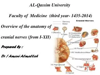
AL-Qassim University Faculty of Medicine (third year- 1435-2014) Overview of the anatomy of cranial nerves (from I-XII) Prepared by : Dr / Amani Almallah
Cranial Nerves: I - XII 12 Pairs of nerves, Numbered from one (I) to twelve (XII). Attach to Ventral surface of brain & exit brain through foramina in skull I + II attach to Forebrain (cerebrum + diencephalon) From III to XII attach to Brainstem 2 attached to mid brain Middle 4: are attached to the pons Lower 4 are attached to the medulla
Functional classification of cranial nerves AFFERENT (SENSORY) EFFERENT (MOTOR) VISCERAL SOMATIC SOMATIC VISCERAL GENERAL VII, IX, X nerves SPECIAL smell and taste (I,VII,IX& X SPECIAL muscles derived from brachial (pharyngeal) arches V, VII , IX and X & XI GENERAL Exteroceptice sensation V GENERAL GENERAL SPECIAL(special sense hearing & vision II &VIII visceral motor nuclei of nerves relayed in parasympathetic ganglions (III, VII, IX & X). muscles of eye and tongue Cranial nerves III, IV, VI, XII.
Functional classification of fibers of cranial nerve General somatic afferent fibers (GSA): transmit exteroceptive and proprioceptive impulses from head and face to somatic sensory nuclei through the trigeminal nerve. Special somatic afferent fibers (SSA): transmit sensory impulses from special sense organs of vision CN II, equilibrium and hearing to the brain(CN VIII). General visceral afferent fibers (GVA): transmit interoceptive impulses from the viscera to the visceral sensory nuclei of VII, IX, X nerves . Special visceral afferent fibers (SVA): transmit sensory impulses from special sense organs of smell and taste to the brain sensation VII, IX, X & smell sensation cranial nerve (CN I).
General somatic efferent fibers (GSE): innervate skeletal muscles of eye and tongue Cranial nerves III, IV, VI, XII. Special visceral efferent fibers (SVE): transmit motor impulses from the brain to skeletal muscles derived from brachial (pharyngeal) arches of embryo. These include the muscles of mastication, facial expression and swallowing Cranial nerves V(1ST arch), VII (2nd arch), IX (3rd arch) and X & XI (cranial part) for (4th& 6th arch). General visceral efferent fibers (GVE): transmit motor impulses from the general visceral motor nuclei and relayed in parasympathetic ganglions. The postganglionic fibers supply cardiac muscles,smooth muscles and gland as Edinger-westphal nuceusof III, superior and inferior salivary of VII, IX respectively, dorsal nucleus of vagus (X).
Olfactory Nerve (= CN I) Functional classification: SVA(special visceral afferent) – purely sensory Carry smell sensation Originated from: Olfactory Epithelium in nasal cavity & filaments passes through cribriform plate of ethmoid. Only CN directly attached to Cerebrum
Optic Nerve Type: Special Somatic Afferent (SSA) Function: Vision Formed by axons of the ganglion cells in the retina. • The nerve passes posteromedially in the orbit, exits through the optic canal to enter the middle cranial fossa, running postero-medially towards the optic chiasma where partial decussation of its fibres occurs. .
Deep origin of III, IV and VI nerves • Functions and nuclei: • The oculomotor: GSE , GVE (Motor nucleus of III & Edinger-Westphal nucleus). • The trochlear: GSE: Motor nucleus of IV. • The abducent: GSE: Motor nucleus of VI IV nucleus VI nucleus VII nucleus III VI IV 7
The Oculomotor nerve Functions: GSE .GVE Deep origin: 1-Motor nucleus of oculomotor (GSE): located in the mid brain, at the level of superior colliculus 2- Edinger-Westphal nucleus (GVE) situated behind main motor nucleus. Axons from the Edinger-Westphal nucleus terminate in the Ciliary ganglion. Postganglionic fibers pass through the short ciliary nerves to the eyeball, where they supply the sphincter pupillae muscle of the iris and the ciliary muscle.
Superficial origin: it emerges on the anterior surface of the midbrain in the interpeduncular fossa, on the medial border of cerebral peduncle & passes forward between the posterior cerebral and superior cerebellar arteries. • In the middle cranial fossa lies in the lateral wall of the cavernous sinus. • As it leaves the sinus, it divides into two divisions, superior and inferior both will enter the orbital cavity through the superior orbital fissure . • Its distribution: • The superior division passes above the optic nerve and ends by supplying: • ▪The superior rectus ▪The levator palpebrae superioris. • The inferior division passes below the optic nerve and ends by supplying: • ▪The inferior rectus ▪ The medial rectus ▪The inferior oblique • ▪ Branch from the nerve to inferior oblique to the ciliary ganglion.
Occulomotor nerve palsy • Lesion results in: • Lateral squint • Ptosis • Diplopia • Pupillary dilatation • Loss of accommodation • Impaired downward & outward movement of the eye ball on the damaged side. The preganglionic parasympathetic fibers run superficially in the nerve and are therefore the first axons to suffer when a nerve is affected by external pressure. Consequently, the first sign of compression of the occulomotor nerve is ipsilateral slowness of the pupillary response to light.
The Trochlear nerve • Function: GSE (purely motor) • Deep origin: The motor nucleus of the trochlear nerve in located in the anterior part of the periaqueductal grey matter at the level of inferior colliculus. • Superficial origin: • From the back of the midbrain below the inferior • colliculus on the sides of the frenulumveli. • The nerve emerges immediately from the • posterior surface of brain stem then passes • on the lateral border of the cerebral peduncle • to passes anteriorly. • It passes through middle cranial fossain the lateral wall of the cavernous sinus below the oculomotor and above the ophthalmic nerves, then enters the orbit through the superior orbital fissure outside the common tendinous ring.
In the orbit it passes medially to enter the orbital surface of the superior oblique muscle (SO 4). • So: • The tochlear nerve the only cranial nerve exits from the posterior aspect of the brain stem. • It supplies superior oblique muscle of the eye ball (SO4)
Trochlear nerve palsyLesion results in diplopia & inability to • rotate the eye infero-laterally. • The eye deviates; upward and • slightly inward. • Person has difficulty in walking downstairs
The Abducent nerve • Function: GSE • Deep origin: • 1- Motor nucleus of the abducent nerve in floor of upper part of 4th ventricle beneath the facial colliculus. • Superficial origin: • The nerve exits At the junction of • the pons and the Upper border of • medulla at the pyramid (pontomedullary angle) • It then, enters the floor of the cavernous sinus • lying lateral to the internal carotid artery. • It enters the orbit through the superior orbital • fissure within the common tendinous ring to end • by supplying the lateral rectus (LR 6).
So The Abducent nerve then enters the orbit through the superior orbital fissure • Abducent nerve palsy • Lesion results in: Medial squint with an inability to • direct the affected eye laterally • A nuclear lesion may also involve the nearby • nucleus or axons of the facial nerve, causing • paralysis of all the ipsilateral facial muscles.
The 3rd, the 4th and the 6th nerve motor nuclei receive: • Corticonuclear fibers from both cerebral hemispheres. • Tectobulbar fibers from superior colliculus. • Fibers from medial longitudinal bundle (MLB) by which the nuclei are connected to each other.
Trigeminal nerve Functions: it is the largest cranial nerve. It has four nuclei one motor & three sensory. It carries motor supply to the muscles of mastication and transmits sensory information from the face, oral and nasal cavities, and most of the scalp. Its function classification: its four nuclei give (GSA &SVE) 1- Motor nucleus supplies: Special visceral efferent (SVE) Its axon form the motor root of trigeminal which join the mandibular nerve. It lies in the middle of pons in line with nucleus amniguus and facial nucleus. It supplies: 1- Muscles of mastication ;temporalis, masseter, medial & lateral pterygoid. 2- Anterior belly of digastric & mylohyoid muscles. 3-Tensor tympani & tensor palati. (muscles of 1st pharyngeal arch)
Nuclei of General somatic afferrent: (GSA) 2- Principal (main) sensory nucleus: it is GSA nucleus, lies in the middle of the pons lateral to the motor nucleus. It receives tactile impulses from face. 3- Mesencephalic nucleus: it is GSA nucleus, called mesencephalic as it extends to the midbrain & junction between (pons & midbrain). It receives proprioceptive fibers from face. 4- Spinal nucleus: it is GSA nucleus. Lies in the lower part the pons, Extending to the whole length of medulla & upper 2-3 cervical segments of spinal cord). It receives pain & temperature sensations from face.
Superficial origin: Emerges From the ventral surface of the pons by a short trunk has small motor root & large general sensory root. • Course and relations: • It is the shortest cranial nerve, its trunk is formed of small motor and large sensory roots The trigeminal ganglion which lies near the apex of the petrous bone on trigeminal impression. • On the surface of the ganglion it divided into its three main divisions • The three main divisions of the trigeminal nerve: • I-The ophthalmic nerve (sensory) • II- The maxillary nerve (sensory) • III- The mandibular nerve (which is joined with the motor root).
Distribution of the function of the trigeminal nerve divisions
The Ophthalmic nerve (V1) • Function: GSA (pure sensory) • Course and relations: It is the smallest of the three branches of the trigeminal. It passes forwards in the lateral wall of the cavernous sinus below the trochlear nerve and above the maxillary nerve. Just before it enters the orbit through the superior orbital fissure, it divides into three branches; lacrimal, frontal & nasociliary. • 1- The lacrimal nerve: • Enters the orbit in the lateral part of the superior orbital fissure it passes along the lateral wall of the orbit. It receives communication (parasympathetic) from the zygomatico-temporal nerve which carries secretory fibres to the lacrimal gland. • It supplies the lacrimal gland, and gives a palpebral branch to the upper eyelid. • 2- The frontal nerve: • It Is the largest branch of the ophthalmic nerve, Enters the orbit through the superior orbital fissure medial to the lacrimal nerve.
In the orbit, the frontal nerve ends by dividing into: • 1-Supratrochlear nerve: to the forehead and the scalp. • 2- Supraorbital nerve: to the frontal air sinus, face and the scalp. • 3-The nasociliary nerve • Enters the orbit through the superior orbital fissure. It ends by giving the following branches: 1- Sensory branch to the ciliary ganglion • 2- Long ciliary nerves : to the eye ball. • 3- Posterior ethmoidal nerve: through the posterior ethmoidal canal to the ethmoidal and sphenoidal air sinuses • 4- Infratrochlear nerve: to the lower eyelid and the face. • 5- Anterior ethmoidal nerve
Summary of ophthalmic nerve It is the smallest branch of the trigeminal. Only sensory Supplies : cornea, conjunctiva, upper lid, forehead anterior part of the scalp & nose. Its course: emerges from trigeminal ganglion lat wall of cavernous sinus Three branches in anterior part of cavernous sinus Lacrimal, Nasociliary & Frontal orbit Superior orbital fissure
The maxillary nerve • Function: GSA (pure sensory) • Course and relations: • It passes in the lateral wall of the cavernous sinus below the ophthalmic nerve. It leaves the cranial cavity through the foramen rotundum to the pterygopalatine fossa, then through the pterygomaxillary fissure to the infratemporal fossa. • It passes through the infraorbital groove and canal in the floor of the orbit, continues as the infraorbital nerve which it appears in the face through the infraorbital foramen. Branches: I- In the cranial cavity: • meningeal branch to the dura matter of middle meningeal fossa • II- In the pterygopalatine fossa: • 1- Ganglionic: sensory branch to the pterygopalatine ganglion • 2- Zygomatic nerve: divides into zygomaticotemporal and zygomaticofacial nerves to the face and scalp (the zygomaticotemporal nerve carries secretory fibers to the
Maxillary nerve Maxillary nerve during its passage through the infra-temporal fossa (to sphenopalatine fossa)
. • lacrimal gland from the ganglion to the lacrimal nerve). • 3- Posterior superior alveolar nerve: pierces the infratemporal surface of the maxilla to supply the maxillary sinus and the molar teeth, it give branches to the cheek and gum. • III- In the infraorbital canal (branches of the infraorbital nerve): • 1- Middle superior alveolar: to the upper premolar teeth • 2- Anterior superior alveolar nerve: to the incisor and canine teeth, it sends a nasal branch to the nose and the nasal septum • IV- Finally; In the face: to supply the face • 1- Palpebral nerve 2- Nasal nerve 3- Superior labial nerve
Summary of maxillary nerve 2nd division of the trigeminal nerve. It is pure sensory. Its course: emerges from trigeminal ganglion lat wall of cavernous sinus To the foramen rotundum To pterygopalatine fossa then in groove on the posterior surface of maxilla • Continues through the infra-orbital fissure in the floor of the orbit as Infra-orbital nerve through the infra-orbital foramen to appears in the face, dividing into its three terminal branches; Palpebral, Nasal & Superior labial nerves.
The mandibular nerve • Functions: GSA, SVE (mixed nerve) • Course and relations: • Is the largest branch of the trigeminal nerve, formed of two roots a large sensory and small motor • It leaves the skull through the foramen ovale to the infratemporal fossa where the two roots unite to form the nerve trunk • The trunk lies medial to the lateral pterygoid muscle • It is very short trunk which divides into anterior and posterior divisions • Branches: • From the trunk then from the anterior & posterior division. • I- Branches From the trunk • 1- Nervus spinosus (sensory, meningeal): it passes up to the cranial cavity through the foramen spinosum to supply the dura matter of the middle cranial fossa.
2- Nerve to medial pterygoid (motor): is the motor to the medial pterygoid muscle, it passes through the otic ganglion without relay then exits to supply the tensor palati and tensor tympani muscles. • II-Branches from the anterior division: (3 motor branches to 3 muscles of mastication +1 sensory): • 1- Deep temporal nerves: to the temporalis muscle • 2- Nerve to lateral pterygoid: supplies lateral pterygoid muscle. • 3- Masseteric nerve: pass through the mandibular notch to masseter. • 4- Buccal nerve (sensory): passes between the two heads of the lateral pterygoid to the face to supply (sensation) to the skin over the cheek. • III- Branches from the posterior division • 1- The auriculotemporal nerve (sensory): • It arises by two roots which surrounds the middle meningeal artery. And unites behind the artery, then it passes deep to the neck of the mandible within the substance of the parotid gland to leave the gland through its upper end of the gland to the scalp.
The auriculotemporal nerve is sensory supply to: • I- auriculotemporal nerve carries postganglionic secretormotor fibres from the otic ganglion & sensory fibers to the parotid gland. • II- skin of upper 2/3 of the lat surface of the auricle & skin of the external acoustic meatus and the outer covering of the ear drum . • III- posterior ½ of temporal area. • IV- atricular branches to Tempro-mandibular joint (TMJ). • 2- The lingual nerve (sensory): • As it originates deep to the lateral pterygoid muscle, it is joined by the chorda tympani branch of the facial nerve in the infratemporal fossa. • Then descends downwards between the medial pterygoid muscle and the ramus of the mandible, then it lies behind the root of the (last) third molar tooth where is covered by the mucous membrane of the mouth (dangerous position).
In the submandibular region, it passes between the mylohyoid laterally and the hyoglossus muscles deep to the submandibular gland. • Termination: The terminal branches of the lingual nerve carries sensations from the mucous membrane of the mouth, the gum, the anterior 2/3 of the tongue. • The chorda tympani through it, supplies the submandibular and the sublingual glands and carries taste fibres from the anterior 2/3 of the tongue. • 3- The inferior alveolar nerve: (Mixed motor & sensory nerve) • It descends deep to the lateral pterygoid, then between the ramus of the mandible and the sphenomandibular ligament to enter the mandibular canal. • It runs in the canal opposite the mental foramen t divides into its terminal branches; mental and incisive branches. • Before it enters the mandibular canal it gives its mylohyoid branch which descends in a groove on the medial side of the ramus of the mandible to end by supplying the mylohyoid and the anterior belly of digastric muscle.
Summary of mandibular nerve It is the largest branch of trigeminal nerve. It is a mixed nerve. It is the nerve of the 1st pharyngeal arch (muscles of mastication). Motor root with the sensory root exit through foramen ovale in greater wing of sphenoid. The trunk of the mandibular lies in the infra-temporal fossa. It is divided into Large posterior division Branches of poterior division: 1- Auricul-temporal nerve (S) 2- Lingau nerve (S) 3- Inferior alveolar nerve( contains sensory & motor fibers). small anterior division Branches of anterior division: 1- Masseteric nerve (M) 2- deep temporal nerve (M) 3- N. to lat pterygoid muscle (M) 4- Buccal nerve (S)
Facial nerve (CN VII) Function of the facial nerve It is a mixed nerve (sensory for the tongue & motor for the face) and sharing in two of H & N parasympathetic ganglia. Motor supply for the muscles of the face (Buccinator , Orbicularis occuli, Orbicular oris, Platysma , Stapedius and etc. Functional classification: Special visceral efferent (SVE) facial muscle s are muscle of the 2nd pharyngeal arch. General visceral efferent (GVE): (sharing in two Parasympathetic ganglia; submandibular ganglion to Submandibular & sublingual & sphenopalatine ganglion to lacrimal ).
Special visceral afferent (SVA ): taste sensation for the anterior 2/3 tongue. General somatic afferent (GSA): (skin over the back of ear) Deep origin: nuclei of facial nerve: 1- Motor nucleus of facial nerve (SVE): lies in the lower part of the pons . 2- Superior salivatory nucleus (GVE): lies in the pons lateral to the main motor nucleus of VII and gives rise to secretomotor parasympathetic fibers that pass in greater superficial petrosal nerve and chorda tympani. 3- Nucleus solitarus (SVA): lies in the medulla, receives the taste sensation from the anterior 2/3 of the tongue via the central processes of the cell of the geniculate ganglion of the facial nerve. 4- GSA fibers : through fibers to acoustic meatus & back of auricle through communication from auricular branch of vagus. This fibers terminate in main sensory nucleus & spinal nucleus of trigeminal Internal course: the motor fibres passes dorsally and medially forming a loop around the abducent nucleus in the floor of the 4th ventricle forming facial colliculus.
FACIAL NERVE • GVE (parasympathetic) from Superior salivary nucleus • SVE from Motor nucleus of VII • GSA & GVA both to Nucleus solitarius
Superficial origin: at the pontomedullary angle above the inferior cerebellar peduncle. • The facial nerve is formed mainly of two parts: • 1- Facial nerve proper (motor): arising from facial motor nucleus in pons. • 2- Nervus intermedius: it is the sensory root of facial lies position between the facial proper and vestibulcochlear nerve in the pontocerebellar angle. Carrying para-sympathetic fibers (from superior salivary nucleus) and taste fibers ( to the solitary nucleus). • Course and relations: • I- Intracranial (intrapetrosal) course II- Extracranial course • I- The intrapetrous course: (LB D) • The nerve passes laterally with the vestibulocochlear nerve (CN VIII) to the internal auditary meatus. At the bottom of the meatus the nerve enters the facial bony canal where it runs Laterally above the vestibule of inner ear. Reaching the medial wall of the middle ear, it bends sharply Backwards above the promontory (forming its genu where the genicular ganglion is found).