Aplastic Anemia
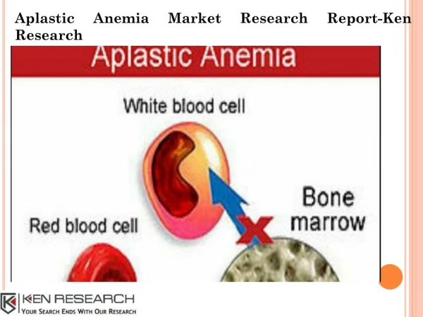
Aplastic Anemia. Andrew J Avery A.M. Report 04/30/10. Introduction. Aplastic anemia is a syndrome of bone marrow failure characterized by peripheral pancytopenia and marrow hypoplasia


Aplastic Anemia
E N D
Presentation Transcript
Aplastic Anemia Andrew J Avery A.M. Report 04/30/10
Introduction • Aplastic anemia is a syndrome of bone marrow failure characterized by peripheral pancytopenia and marrow hypoplasia • Pancytopenia is a reduction in the peripheral blood of all three cellular components (i.e. anemia, neutropenia and thrombocytopenia)
Introduction • Paul Ehrlich introduced the concept of aplastic anemia in 1888 when he studied the case of a pregnant woman who died of bone marrow failure • In 1904 AnatoleChauffard named this disorder aplastic anemia
Pathophysiology • Complicated and beyond the scope of this presentation, but it is felt that 80% of cases of aplastic anemia are acquired • It can be difficult to distinguish primary vs acquired aplastic anemia • In acquired aplastic anemia, clinical and laboratory observations suggest that this is an autoimmune disease. • Supported by the finding that ≈ 70% of pts with acquired aplastic anemia impove with immunosuppressive therapy
Epidemiology • Several retrospective studies suggest that the incidence is 0.6-6.1 cases per million population in the United States • Incidence is much more common in Asia: 4 cases per million in Bangkok, and as high as 14 cases per million in Japan (likely 2/2 environmental factors, as an increased frequency is not seen in persons of Asian ancestry living in the US)
Epidemiology • Male to Female ratio is 1:1 • Occurs in all age groups: small peak in childhood 2/2 inherited marrow-failure syndromes; 2nd peak in people aged 20-25 years, and a subsequent peak is observed in people older than 60 years (this 3rd peak may be related to inclusion of MDSs, which are unrelated to aplastic anemia)
Diagnostic Criteria • Moderate aplastic anemia — The criteria for moderate AA include: • Bone marrow cellularity <30% • Absence of severe pancytopenia • Depression of at least two of three blood elements below normal
Diagnostic Criteria • Severe aplastic anemia — The criteria for severe aplastic anemia (SAA) are: • A bone marrow biopsy showing <25% of normal cellularity, or • A bone marrow biopsy showing <50% normal cellularity in which fewer than 30% of the cells are hematopoietic and at least two of the following are present: absolute reticulocyte count <40,000/microliter; ANC <500/µL; or plt count <20,000/µL.
Diagnostic Criteria • Very severe aplastic anemia — The patient is considered to have very severe aplastic anemia (vSAA) if the criteria for severe aplastic anemia are met and the ANC is <200/µL
Clinical Manifestations • The onset of sxs is insidious, and the initial symptoms are related to anemia or bleeding, although fever or infections are also often noted at presentation • Anemia may manifest as pallor, headache, palpitations, dyspnea, fatigue, or foot swelling • Thrombocytopenia may result in mucosal and gingival bleeding or petechial rashes
Clinical Manifestations • Neutropenia may manifest as overt infections, recurrent infections, or mouth and pharyngeal ulcerations
History and Physical Exam • A detailed work history, with emphasis on solvent and radiation exposure should be obtained, as should a family, environmental, travel, and infectious disease history • Exam may show signs of anemia, such as pallor and tachycardia, and signs of thrombocytopenia, such as petechiae, purpura, or ecchymoses. Overt signs of infection are usually not apparent at diagnosis
Physical Exam • A subset of patients with aplastic anemia present with jaundice and evidence of clinical hepatitis • Adenopathy or organomegaly should suggest an alternative diagnosis (eg lymphoma or leukemia) • Look for physical stigmata of inherited marrow-failure syndromes, such as skin pigmentation, short stature, microcephaly, hypogonadism, mental retardation, and skeletal anomalies
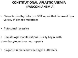
Causes • Congenital or inherited causes of aplastic anemia (20%) • Patients usually have dysmorphic features or physical stigmata. On occasion, marrow failure may be the initial presenting feature • Fanconi anemia • Dyskeratosiscongenita • Cartilage-hair hypoplasia • Pearson syndrome • Amegakaryocytic thrombocytopenia (thrombocytopenia-absent radius [TAR] syndrome)
Congenital or Inherited Causes • Shwachman-Diamond syndrome • Dubowitz syndrome • Diamond-Blackfan syndrome • Familial aplastic anemia
Causes • Acquired causes of aplastic anemia (80%) • Idiopathic factors • Infectious causesz: Hepatitis Viruses, EBV, HIV, Parvovirus, and Mycobacteria • Toxic Chemical: Benzene, Lindane, Glue Vapers, and Radiation • Idiosyncratic Drug Rxns: Chloramphenicol, Gold, NSAID (phenylbutazone,indomethacin), Sulfonamides, AEDs (felbamate), Arsenicals
Acquired Causes • Immune Disorders: SLE, GVHD, Eosiniphilic Fasciitis • Miscellaneous: Paroxysmal Nocturnal Hemoglobinuria, Thymoma, Thymic carcinoma, and Pregnancy
Differential Diagnosis • ALL, MDS, AML, Myelophthisic Anemia, Agnogenic Myeloid Metaplasia With Myelofibrosis, Osteopetrosis, HHV 6, SLE, Non-Hodgkins Lymphoma, Megaloblastic Anemia, and Multiple Myeloma
Workup • Laboratory Studies: • CBC w/diff: will show pancytopenia, a reduction in the absolute number of reticulocytes, and possibly mild macrocytosis • Peripheral Blood Smear: helpful in distinguishing aplasia from infiltrative and dysplastic causes -
Workup • Bone Marrow Bx: • The bone marrow is profoundly hypocellular with a decrease in all elements; the marrow space is composed mostly of fat cells and marrow stroma • Infiltration of the bone marrow with malignant cells or fibrosis is not present • Residual hematopoietic cells are morphologically normal and hematopoiesis is not megaloblastic
Additional Tests • Hemoglobin electrophoresis and blood-group testing: may show elevated levels fetal hemoglobin and red cell I antigen, suggesting stress erythropoiesis (found in MDS & AA) • Serologic Testing for Viral Entities • Measurement of red cell membrane CD59 if PNH is considered (better than HAM test) • Diepoxybutane incubation is performed to assess chromosomal breakage for Fanconi anemia • An eval for autoimmune collagen-vascular dz
Treatment • Treatment of AA includes withdrawal of potentially offending agents, supportive care (eg, transfusion, antibiotics), and some form of definitive therapy (eg, hematopoietic cell transplantation, immunosuppressive regimens). Blood and platelet transfusions should be used selectively in patients who are candidates for HCT to avoid sensitization
Treatment • HCT: Allogeneic hematopoietic cell transplantation (HCT) is curative in AA, but is limited by the availability of an HLA-matched sibling as well as by the potentially fatal consequences of graft versus host disease in patients over the age of 40 to 45 • Immunosuppressive regimens: Immunosuppressive regimens are not curative, but can be associated with long-term survival
Prognosis • The prognosis of aplastic anemia (AA) depends upon two factors, disease severity and patient age • Effect of age — There is a strong inverse relation between patient age and five-year survival in patients with AA • Unless patients with SAA or vSAA are successfully treated, over 70% will be dead within one year. At any degree of severity of AA, the outcome is worse in older patients