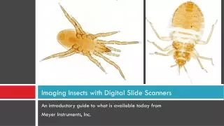
Imaging Insects with Digital Slide Scanners
320 likes | 657 Vues
Imaging Insects with Digital Slide Scanners. An introductory guide to what is available today from Meyer Instruments, Inc. Scanning History.

Imaging Insects with Digital Slide Scanners
E N D
Presentation Transcript
Imaging Insects with Digital Slide Scanners An introductory guide to what is available today from Meyer Instruments, Inc.
Scanning History The first digital slide scanner based on film scanner technology was designed for Pathology. The intended use was to image and share tissue from whole histological 1x3” glass slides. • Basic instrument consisted of a modified 35mm film scanner and a modified slide holder • Meyer Instruments, Inc. was the first to patent, and the product the PathScan Enabler was launched in 1995
Scanning History The first digital slide scanner based on microscope technology was developed in early 1975. The intended use was to image cells from histological 1x3” glass slides. • Primary use was to facilitate and enable high speed image acquisition of cells for clinical purposes • The product was never fully implemented or marketed
Honeywell’s 1975 patent “A scanning microscope system with automatic cell-find and autofocus”
Scanning History Subsequent microscope technology based digital slide scanners were developed in early 2001. The intended use was to capture high resolution images from histological 1x3” glass slides for pathology use. • Basic instrument consisted of a research grade microscope with motorized X,Y and Z control, a camera, stitching software and a computer workstation • Most were big, cumbersome and very expensive
Interscope 2001 patent “A microscope based design for pathology use’’
Scanning History Numerous companies followed with other microscope designs, including: Hamamatsu, Aperio, Zeiss, Leica, Nikon, Olympus, Ventana, GE, Mikron, Perkin Elmer, Sakura, Huron, Motic, Objective Imaging and others Most are only capable of scanning one plane of focus (Z focus is not possible)
Imaging Insects with Scanners Recommended requirements: • Scanner with XY and Z focus control • Extended depth of field (EDF) software • Enhancement software
Imaging Insects with Scanners Scanner companies with XYZ imaging control to consider include: Motic, Objective Imaging and Hamamatsu
Imaging Insects with Scanners Companies with Extended Depth of Field (EDF) software to consider include: Media Cybernetics www.mediacy.com Motic www.moticusa.com Huvitz www.microscope.huvitz.com HeliconSoft www.heliconsoft.com Zerene Systems www.zerenesystems.com
Imaging Insects with Scanners Companies with Image Enhancement Software to consider include: Adobe Photoshop www.adobe.com Media Cybernetics www.mediacy.com ImageJ www.imagej.nih.gov/ij/index.html
Advantage of Scanners Compared to conventional microscope-camera based photography: Entire insect can be captured at higher resolution Insect is entirely in focus Larger, entirely in focus insects can be printed on large format printers for stunning posters
Conventional microscope-camera photography allow only a single frame with limited field of view
Scanners create mosaic images in XY & Z resulting in an overall superior picture
Sample images using the Motic VM600 digital slide scanner Helicon, Image-Pro Plus and Photoshop
Sample images using the Motic VM600 digital slide scanner Helicon, Image-Pro Plus and Photoshop
Sample images using the Motic VM600 digital slide scanner Helicon, Image-Pro Plus and Photoshop
Sample images using the Motic VM600 digital slide scanner, Helicon, Image-Pro Plus and Photoshop
Sample images comparing 3 different HeliconSoft EDF image algorithm’s
Sample images comparing using 40x objective verses 20x objective
Final sample image using 40x objective and the Motic VM600 scanner with HeliconSoft EDF software
Lets look live at the Motic VM600 • Also using Helicon Soft • Also using Photoshop • Also using Image-Pro Plus Advantages over normal microscope photography • Entire insect can be captured at higher resolution • Insect is entirely in focus • Larger, entirely in focus insect can be ‘jumbo’ printed
Motic VM600 Slide Scanner • Microscope based design • Least expensive XYZ slide scanner on the market • Scans with 2x, 4x, 10x, 20x, 40x objective's • Comes complete with computer imaging work station • Many optional features include distance remote control • Files can be exported in a variety of image formats • Easy set up and use
Objective Imaging Scanner • Microscope in a box based design • Scans with single 20x high n.a. objective • Integrated computer with touch screen makes for very compact footprint • Fast two slide scanning capacity • Built-in extended depth of field (EDF) software • Files can be exported in a variety of image formats
Hamamatsu Slide Scanners • Microscope in a box, true tri-linear array based design • High performance XYZ walk away batch scanning • Scans with single 20x high n.a. objective • Comes complete with computer imaging work station • High through put 6, 210, or 320 slide capacity • Z scans for up to 2mm thick samples • Fluorescence optional available on all models
To image insects with scanners • You must have a XYZ slide scanner • You must have EDF software • You must have Enhancement software • You must have a powerful computer • You must have professional training
To conclude, most slide scanners • Are now a “microscope in a box design” • Include a single 20x objective • Do not allow Z focus stacks • Allow multiple slide capacity • Designed primarily to scan histology slides • Are not intended to scan thick samples • Will not (necessarily) scan insects well
Meyer Instruments, Inc. • Independent microscope dealer for 28 years • Distributor of digital slide scanners including PathScan Enabler, Hamamatsu, Mikron, Objective Imaging, Motic • Designed and patented PathScan Enabler • Developed Extended Depth of Field (EDF) algorithm for Media Cybernetics • Created Realtime Telepathology Imaging System (RTIS)
1304 Langham Creek Dr., Suite 235 Houston, TX 77084 (281) 579-0342 www.meyerinst.comwww.realtimetelepathology.com