Download
1 / 16
180 likes | 464 Vues
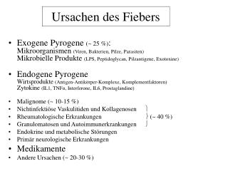
Ursachen des Fiebers. Exogene Pyrogene ( ~ 25 %) : Mikroorganismen (Viren, Bakterien, Pilze, Parasiten) Mikrobielle Produkte (LPS, Peptidoglycan, Pilzantigene, Exotoxine)

E N D
Ursachen des Fiebers • Exogene Pyrogene (~ 25 %):Mikroorganismen (Viren, Bakterien, Pilze, Parasiten)Mikrobielle Produkte (LPS, Peptidoglycan, Pilzantigene, Exotoxine) • Endogene PyrogeneWirtsprodukte (Antigen-Antikörper-Komplexe, Komplementfaktoren)Zytokine (IL1, TNFα, Interferone, IL6, Prostaglandine) • Malignome (~ 10-15 %) • Nichtinfektiöse Vaskulitiden und Kollagenosen • Rheumatologische Erkrankungen (~ 40 %) • Granulomatosen und Autoimmunerkrankungen • Endokrine und metabolische Störungen • Primär neurologische Erkrankungen • Medikamente • Andere Ursachen (~ 20-30 %)
Infektionen bei erworbener AbwehrschwächeNeutropenie(< 500/mm3 oder Funktionsstörung) Staphylococcus aureusStaphylococcus epidermidisStreptokokken PneumokokkenPseudomonas aeruginosaEnterobakterienCandida spp.Aspergillus spp. • KM-Transplantation • akute Leukämie • Zytostatikatherapie • Azidose • Verbrennung ≤ 5 Tage → geringes Risiko Neutropeniedauer 6-9 Tage → Standardrisiko ≥ 10 Tage → hohes Risiko
Infektionen bei erworbener Abwehrschwäche:T-Zell-Defekt • Transplantation(Organe oder KM) • lymphatische Leukämie • Morb. Hodgkin • Zytostatikatherapie • Kortisontherapie • HIV-Infektion • Urämie Salmonella spp.Mycobacterium tuberculosisListeria monozytogenes Legionella pneumophila MOTT Nocardia spp.Cryptococcus neoformansToxoplasma gondiiPneumocystis jiroveciCMV, EBV, VZV, HSV-1, -2, -6, -8 Adenoviren Papovaviren (JC-, BK-Virus)
Infektionen bei erworbener AbwehrschwächeAntikörpermangel (quantitativ oder funktionell) • multiples Myelom • lymphatische Leukämie • myeloische Leukämie • Hypogammaglubulinämie • HIV-Infektion Salmonella spp.Mycobacterium tuberculosisListeria monozytogenes Legionella pneumophila MOTT Nocardia spp.Candida spp. Cryptococcus neoformansToxoplasma gondiiPneumocystis jiroveciCMV, EBV, VZV, HSV-1, -2, -6, -8 Adenoviren Papovaviren (JC-, BK-Virus) Pneumokokken Haemophilus influenzae Enteroviren
Infektion nach Splenektomie • Schwere Infektion oder Sepsis mit:- Pneumokokken (OPSI*-Syndrom)- Haemophilus influenzae- Neisseria meningitidis • Malaria • Schwere Verläufe von Infektionen nach Bißverletzung (Capnocytophaga canimorsus) * overwhelming post splenectomia infection
Infektionen bei HIV-Infektion • > 500 CD4-Zellen: recurrierende Candidiasis • 200 – 500 CD4-Zellen: Lungentuberkulose, Zoster, EBV-Infektion, HHV8-Infektion, Candidiasis • < 200 CD4-Zellen: Pneumozystose, Candidiasis, mukokutaner Herpes, Miliartuberkulose, Kryptposporidiose, Mikrosporidiose • < 100 CD4-Zellen: Zerebrale Toxoplasmose, Lymphome, Kryptokokkose, primäre ZNS-Lymphome, Leukenzephalopathie • < 50 CD4-Zellen: CMV-Retinitis, disseminierte M. avium/intracellulare-Infektionen
Mikrobiologische Diagnostik • Initial: Blutkultur, Identifikation, AB-Resistenz • nach Bedarf: Urinkultur Stuhlkultur, Toxin-Nachweis, Virus-Nachweis Haut-, Schleimhautabstriche Liquorzytologie, -Kultur Kultur aus Gewebepunktaten BAL: Aufarbeitung bakteriologisch, virologisch, mykologisch(immer quantitativ!)Venen-Katheterspitzen • Antigennachweise: Candida, Aspergillus, Cryptococcus aus Serum Legionella, Pneumokokken aus Urin pp65 aus EDTA-Blut • PCR ?
Blutkultur - Hauptfehler Kontamination: - Entnahme aus einem Venenkatheter (Kontaminationsrate > 9%!) - unsterile Handschuhe - kein Mundschutz - unzureichende Desinfektion (Haut, Flaschenstopfen) Falsch negatives - laufende Antibiotika-TherapieErgebnis - lange Zwischenlagerung (auch bei 37°C) - kalte Flasche (Entnahme und Transport)
Bakteriologische Diagnostik • Kultur aus Abstrichen, Punktaten, Geweben, BK • Biochemische Typisierung • Serotypisierung • Genotypisierung • Resistenzbestimmung • Staphylococcus aureusStaphylococcus epidermidis • StreptokokkenPneumokokken • Haemophilus influenzae • EnterobakterienSalmonella spp. • Pseudomonas aeruginosa • Legionella pneumophila • Listeria monozytogenes • Mycobacterium tuberculosis • MOTT • Nocardia spp. Antigennachweise aus Urin(Pneumokokken, Legionellen) Nachweis nicht kultivierbarer Bakterien16(23)S r-RNA-Gen-PCR, spez. PCR Klonale ZuordnungenPulsfeld-Gelelektrophorese SPA-Typisierung
p <0.001 p <0.001 Inadäquate Antibiotikatherapie, n=169 Adäquate Antibiotikatherapie, n=486 Inadäquate Antibiotikatherapie ist ein Risikofaktor für die Krankenhausletalität 60 50 40 Krankenhausletalität (%) 30 20 10 0 alle Ursachen Sepsis Kollef MH et al. Chest; 1999 115:462-474
Pilzdiagnostik • Candida spp. • Aspergillus spp. • Cryptococcus neoformans • Pneumocystis jiroveci • Mikroskopie • Erregeranzucht und Typisierung • Antigen-Nachweis Candida (nicht alle Spezies) Aspergillus Cryptococcus • PCR (Serum/Plasma/Liquor) • Serologie
Virus-Nachweis Antikörper-Nachweis CMV-Pneumonie CMV-Nephritis CMV-Hepatitis CMV-Enterocolitis CMV-Retinitis CMV-Enzephalitis CMV: Diagnostik Antigenämie (pp65)*kommerziell DNA-Hybridisierung* PCR aus Leukozyten* PCR aus Plasma*(Viruslast) kommerziell RT-PCR (mRNA/pp67)*kommerziell Virus-Anzucht (shell vial) Resistenztestung phänotypisch genotypisch KBR (obsolet) Neutralisationsteste ELISA, IFT, BlotIgG IgM IgA Avidität AK gegen spezielle AG CMV-spezifische T-Lymphozyten * Nur quantitative Aussagen sind verwertbar!
EBV- Diagnostik B-Zell-Lymphome Haarleukoplakie EBV-assoziierte Tumoren EBV-Hepatitis • Serologie: Immunoblot • mit Detektion von AK gegen verschiedene EBV-spezifische Antigene • Hybridisierung von EBV-RNA in Lymphomen • EBV-PCR
VZV Diagnostik • besonders bei GvHD: • Generalisierte Primärinfektion • Enzephalitis • Pneumonie • Schwerere Zoster • Virus-Anzucht • direkte IF • VZV-PCR • Serologie
HSV-1, -2, -6, -8 - Diagnostik schwere lokale Exazerbationen Enzephalitis Retinitis Pneumonie Hepatitis Virus-Anzucht direkte IF HSV-PCR Serologie:ELISA, Blot
Virologische Diagnostik Pneumonie KCE Zystitis Virus-Anzucht Virus-PCR, Typisierung Antigen-EIA Serologie ? • Adenoviren • Polyomaviren (JC-, BK-Virus) • Parvovirus B19 Pneumonie Nephritis Virus-PCR Anämie Aplastische Krise Arthritis Virus-PCR Serologie: ELISA