Download
1 / 42
510 likes | 802 Vues
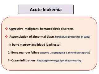
Acute leukemia. Aggressive malignant hematopoietic disorders A ccumulation of abnormal blasts ( I mmature precursors of WBC) in bone marrow and blood leading to: 1- Bone marrow failure ( anemia ,neutropenia & thrombocytopenia)

E N D
Acute leukemia • Aggressive malignant hematopoietic disorders • Accumulation of abnormal blasts (Immature precursors of WBC)in bone marrow and blood leading to: • 1- Bone marrow failure(anemia ,neutropenia & thrombocytopenia) • 2- Organ infiltration ( hepatosplenomegy ,lymphadenopathy )
HISTORY • Means “white blood” in Greek. • Named by pathologist Virchow in 1845. • Classified by FAB classification systems in 1976. • Reclassified by World Health Organization in 2001 & 2008.
Genetic predisposition Environmental factors Infection Previous therapy Other hematological disorders Unknown Mechanism Genetic alteration in the immature precursors Block of differentiation ,Enhanced proliferation & Decreased apoptosis
PATHOGENESIS AML ALL No differentiation Proliferation
Epidemiology • AL represent about 8% of neoplastic disease & cause about 4% of malignancy related deaths ! • AML has an incidence of 2 – 3 per 100 000 per year in children, rising to 15 per 100 000 in adults. • ALL has an incidence of 30 per million & represent about 76% of childhood leukemia .
General Classification Acute leukemia Acute Myeloid Leukemia Acute Lymphoid Leukemia Acute Leukemia of Ambiguous Lineage
Basis of Classification Clinical history (Previous therapy) Morphology Flow cytometry Chromosomal Karyotyping Molecular study
1- Light microscopy(blood smear, bone marrow aspirate & biopsy ) • Blast count : it should be >20% out of the total cells • Blast morphology : Myeloblast: -Size: medium-Large -Nucleous: round, oval or irregular -Nucleolus: prominent -Cytoplasm: abundant, granular Auer rods is characteristic • Lymphoblast: • Size: small- medium • Nucleous: round • Nucleolus: not prominent • Cytoplasm: scanty ,agranular • may be vacuolated
2-Flow cytometry: Laser based technology allows for cells counting & detection of their surface &cytoplasmic markers by suspending them in a stream of fluid followed by analysis through electronic system.
Basis of Classification Stem Cell Markers: (CD34& TDT) Myeloid B-Lymphoid T-Lymphoid MPO CD13 CD33 CD14 CD64 CD41 CD235a CD3 CD4 CD5 CD7 CD8 CD10 CD19 CD22 CD79a
3-Chromosomal Karyotype Set of the chromosomes from one cell during metaphase to study the numerical(deletion &trisomy) and structural ( translation &inversion ) abnormality
4- Molecular studies: Several techniques used to detect and localize the presence or absence of specific DNA sequences on chromosomes Fluorescent In-Situ Hybridization (FISH) Polymerase Chain Reaction (PCR)
Recurrent genetic abnormalities AML ALL
Acute Myeloid Leukemia • Group of hematopoietic neoplasms caused by proliferation of malignant myeloid blasts in bone marrow and blood. • The blast ≥20% or t(8;21) t (16;16) or t(15;17). • More in Adults (do occur in infants!) • Worse than ALL
HSC Megakaryoblast Erythroblast
Mixed M0 ,M1 HSC M4 M7 M6 M5 M2 Megakaryoblast Erythroblast M3 B-ALL T-ALL
FAB Classification • Based on morphology& flow cytometry CD235a CD41
AML Classification (WHO) Myelodysplasia related AML Therapy related AML AML, not otherwise specified (FAB) AML with recurrent genetic abnormalities 1- t(8;21) 2- t(16;16) 3- t(15;17) Prognosis: Good • Blasts≥ 20% • Significant dysplasia • Prognosis: • poor • Blasts≥ 20% • Previous chemotherapy • Prognosis: • poor • Blasts≥ 20% • Genetic: N • No dysplasia • Prognosis: • Standard
M0 M1 M3 M2
M5 M4 M6 M7
Clinical Features of AML 1-Pancytopenia: • WBC infection (fever ,septic shock) • Hb anemia (fatigue , headache , pallor ,SOB….) • platelets bleeding (bruises , epistaxis ,menorrhagia…) Acute onset 2-Organ infiltration: • Hepatosplenomegally. • Lymphadenopathy (rare) • Myeloid sarcoma • Gum hypertrophy • CNS disease More with Acute Monoblastic Leukemia
Clinical Features of AML 3-Leucostasis (increased blood viscosity) 4-Disseminated Intravascular Coagulation (DIC): Widespread activation of coagulation system leading to intravascular fibrin deposition &consumption of platelet and coagulation factors which can be manifested as bleeding (85%) or thrombosis (15%) More with Acute Promyelocytic leukemia (M3)
Clinical Features of AML • Myeloid sarcoma • Gum hypertrophy
Case Study • 65 years old male presented to ER with fatigue ,fever and nose bleeding for 2 weeks. • O/E : moderate hepatosplenomegaly & multiple bruises. • CBC : WBC :40 x109/L HB: 7g/dL PLT: 51 x109/L
Blood smear & bone marrow: ? Flow cytometry : The blast are positive for CD34 ,CD13,CD33,CD117 and MPO They are negative for CD3,CD10,CD19&CD79a
Karyotype : The final diagnosis: AML with t(8;21) (WHO)
Prognosis and treatment • Better prognosis: • Genetics: t(8;21), inv(16;16) or t(15;17) • Age: < 60 years • Primary better than secondary • Treatment • Chemotherapy: • AML: M0-M8 but not M3 ( same protocol) • AML: M3 (ATRA or arsenic) • Stem cell transplantation
Acute Lymphoblastic Leukemia (ALL) • Acute leukemia characterized by proliferation of malignant lymphoid blasts in bone marrow and blood. • B and T cells • More common in Children • Better than AML
Clinical Features of ALL 1-Pancytopenia: • WBC infection (fever ,septic shock) • Hb anemia (fatigue , headache , pallor ,SOB….) • platelets bleeding (bruises , epistaxis ,menorrhagia…) Acute onset 2-Organ infiltration: • Lymphadenopathy (very common) • Hepatosplenomegally. • testicles involvement • CNS disease • Mediastinal mass Characteristic for T-ALL
L3 (Burkitt’s) represents mature lymphoid neoplasmso it is a type of lymphoma not Acute lymphoblastic leukaemia
B-ALL Precursor B cell Mature B cell CD34& TDT Surface Immunoglobulin CD10 Common B-ALL CD19,CD20 &CD79a B- ALL Burkitt’s
T-ALL Precursor T- cell Mature T- cell cCD3 sCD3 - VE +VE CD4 only (CD4&CD8) (CD4&CD8) CD8 only CD2,CD5&CD7 T-ALL T- Cell Lymphoma 38
Prognosis &treatment • Treatment: • Chemotherapy (high cure rate) • Stem cell transplantation
Remember ! • Acute leukaemia is a fatal neoplastic condition • 20% or more blasts = Acute leukaemia • Diagnosis requires special investigations • Auer rods = AML • AML M3 = DIC &target therapy • Gum hypertrophy = mostly M4 or M5, • Mediastinal = T-ALL
Remember ! • Subtypes of AML (M0-M8) + cytogenetic abnormalities • Subtypes of ALL (T or B cell) • Main lineages markers are MPO, CD19 and CD3 • Stem cell markers are CD34,TDT • FAB classification based mainly on morphology • WHO classification focused more on genetics