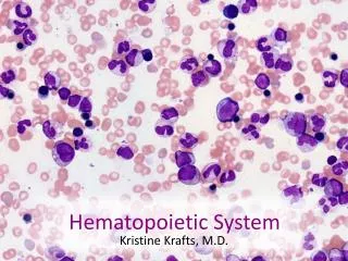
Hematopoietic System
Hematopoietic System. Dr. Mohamad Nidal Khabaz Assistant Professor of Pathology, Pathology Department, Faculty of Medicine, Jordan University of Science and Technology. Hematopoiesis. All mature blood cells originate from pluri-potent stem cells in the bone marrow.

Hematopoietic System
E N D
Presentation Transcript
Hematopoietic System Dr. Mohamad Nidal Khabaz Assistant Professor of Pathology, Pathology Department, Faculty of Medicine, Jordan University of Science and Technology
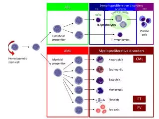
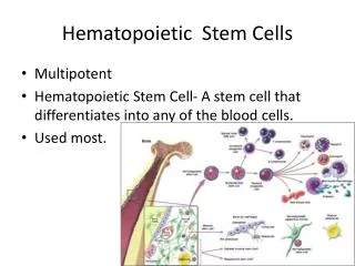
Hematopoiesis • All mature blood cells originate from pluri-potent stem cells in the bone marrow. • Pluripotent stem cells have high level of proliferation and self reproduction. They may change through several steps of development to different cell types. • Maturation of cells is going through several intermediate steps. • Usually only mature cells can reach blood
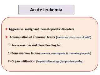
Normal bone marrow. Note the presence of megakaryocytes, erythroid islands, and granulocytic precursors.
Hematopoiesis • Capacity to differentiate is increasing and capacity to divide and sel-production is decreasing during maturation. Mature blood cells are not able to divide. • The proliferation, differentiation, and functional abilities of the various blood cells are controlled by cytokines (produced by lymphocytes and stromal cells of the bone marrow). • Hematopoiesis must and is very carefully regulated to hold stable number of cells in the blood.
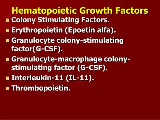
Hematopoiesis • Cytokines (colony stimulating factors) stimulate hematopoietic cell colonies from bone marrow precursors: • EPO (Erythropoietin) • TPO (Thrombopoietin) • G-CSF (Granulocyte colony stimulating factor) • GM-CSF (Granulocyte-monocyte colony stimulating factor) • Increase peripheral stem cells for transplantation • Accelerate cell proliferation after bone marrow engraftment
HEMATOPOIETIC FACTORS n, neutrophils; m, monocytes; e, eosinophils; b, basophils; meg, megakaryocytes; rbc, red blood cells IL – interleukin , CSF – colony stimulating factor
Physical Characteristics and Volume of the Blood • Blood is a sticky, thick fluid with a metallic taste • Color varies from scarlet (oxygen-rich) to dark red (oxygen-poor) • The pH of blood is 7.35–7.45 • Temperature is 38C, slightly higher than “normal” body temperature • Blood accounts for approximately 8% of body weight • Average volume of blood is 5–6 L for males, and 4–5 L for females.
Functions of the Blood • Carries oxygen from lungs to all body cells & removes carbon dioxide from the cells. • Carries waste products of cell activity to kidneys to be removed from the body. • Transports nutrients from digestive system to body cells.
Functions of the Blood • Transports hormones from endocrine glands to target organs • Blood cells help fight infection and heal wounds (white blood cells & platelets). • Blood maintains appropriate body temperature by absorbing and distributing heat.
Composition of Blood • Blood separates into two main parts: • Plasma accounts for 55% • Formedelements (Blood cells) 45% of blood volume, Produced in red bone marrow. They are three kinds: • Red = erythrocytes • White = leukocytes • Platelets = thrombocytes
Function of the Plasma • Carry the cells that transport gases • Aid in body defenses • Prevent blood loss
Plasma Proteins • Albumin • Comprise approximately 54% of the plasma proteins • Contribute to plasma osmotic pressure and the maintenance of blood volume • Serve as a carrier for certain substances • Globulins • Comprise approximately 38% of plasma proteins
Plasma Proteins (cont.) • Alpha globulins transport bilirubin and steroids • Beta globulins transport iron and copper • Gamma globulins constitute the antibodies of the immune system. • Fibrinogen • Make up approximately 7% of the plasma proteins • Is converted to fibrin in the clotting process
Blood cells • Red blood cells • Platelets • Leukocytes are 5 types of white blood cells protect against disease: • Basophils. • Eosinophils. • Neutrophils. • Lymphocytes. • Monocytes
Erythrocytes: Red Blood Cells • Normally there are 4 to 6 million RBCs per mm3 of whole blood (Most numerous of blood cells). • Shape: Biconcave discs, 8 micrometers in diameter. • Red blood cells lack nucleus, ER, & mitochondria. • Production regulated by erythropoietin (hormone made in kidneys) • The kidneys produce the hormone erythropoietinto increase blood cell production when oxygen levels are low.
Function of the Red Blood Cells • Transportation of oxygen to the tissues • Hemoglobin binds some carbon dioxide and carries it from the tissues to the lungs • The hemoglobin molecule is composed of two pairs of structurally different polypeptide chains • Each of the four polypeptide chains consists of a globin (protein) portion and heme unit, which surrounds an atom of iron that binds oxygen. • Each molecule of hemoglobin can carry four molecules of oxygen. • The production of each type of globin chain is controlled by individual structural genes with five different gene loci. • Mutations can occur anywhere in these five loci
Erythropoiesis • Red cells are produced in the red bone marrow after birth • Until age 5 years, almost all bones produce red cells to meet growth needs; after 5 years, bone marrow activity gradually declines • After 20 years, red cell production takes place mainly in the membranous bones of the vertebrae, sternum, ribs, and pelvis • With this reduction in activity, the red bone marrow is replaced with fatty yellow bone marrow
Red Blood Cell Destruction • The red blood cell has a life span of approximately 120 days, so requires constant replacement. • It is broken down in the spleen. • Iron is reused by the red bone marrow where stem cells continually produce more red blood cells. • The heme molecule is converted to bilirubin and transported to the liver. • It is removed and turn into water soluble for elimination in the bile. • Lack of enough hemoglobin results in anemia.
Laboratory Tests for Red Blood Cells • Red blood cell count (RBC) • Measures the total number of red blood cells in 1 mm3 of blood. • Percentage of reticulocytes (normally approximately 1%) • Provides an index of the rate of red cell production. • Hemoglobin (grams per 100 mL of blood) • Measures the hemoglobin content of the blood. • Hematocrit • Measures the volume of red cell mass in 100 ml of plasma volume
C.B.C • Haemoglobin - 15±2.5, 14 ±2.5 - g/dl • PCV - 0.47 ±0.07, 0.42 ±0.05 - l/l (%) • Haematocrit, effective RBC volume - better • RBC count - 5.5 ±1, 4.8 ± 1 x10¹²/l • MCHC - Hb/PCV - 30-36 - g/dl • Hb synthesis within RBC • MCH - Hb/RBC - 29.5 ± 2.5 pg/l • Average Hb in RBC • MCV - PCV/RBC 85 ± 8 – fl • Average volume of red cells
Anemia • Definition • An abnormally low number of circulating red blood cells or level of hemoglobin, or both • Low Hb <13.5 (males), <11.5 (females) • Results in diminished oxygen-carrying capacity. • Causes • Excessive loss or destruction of red blood cells • Deficient red blood cell production because of a lack of nutritional elements or bone marrow failure.
Anemia • Manifestations of Anemia • Impaired oxygen transport with the resulting compensatory mechanisms • Reduction in red cell indices and hemoglobin levels • Signs and symptoms associated with the pathologic process that is causing the anemia • fatigue, paleness, shortness of breath, and chills
Decreased production Nutritional Marrow suppression Marrow infiltration Increased loss Blood loss Hemolytic Immune Non immune Acquired: Decreased production Increased loss Congenital: Increased loss/Hemolytic Anemia Classification
Acquired RBC disorders • Decreased Production: • Aplastic, Hypoplastic anemias • Deficiency anemias Iron, B12, Folate etc. • Lack of erythropoiesis - Kidney disease • Marrow disease, malignancy, radiation • Increased loss/destruction: • Blood loss anemias - parasites, bleeding • Hemolytic anemias - Autoimmune (cold & warm antibody) mechanical, drugs & toxins.
Hemolytic Anemias • Inherited disorders • Membrane Disorders: • Spherocytosis, Elliptocytosis • Enzyme disorders: • Glucose-6-phosphate dehyrogenase and pyruvate kinase deficiencies. • Immune lysis (Warm & Cold Ab types). • Acquired hemolytic anemias: Infection induced (Clostridia, malaria, septicemia).
Hemolytic Anemias (Cont.) • Hemoglobinopathies • Sickle cell anemia (the presence of crescent- or sickle-shaped erythrocytes and accelerated hemolysis) • Thalassemia Syndromes , , (inherited disorders of hemoglobin metabolism in which there is a decrease in net synthesis of a particular globin chain without change in the structure of that chain).
Anemias of Deficient Red Cell Production • Iron deficiency anemia (NormalSerum iron (1mg/l)). • Megaloblastic anemias • Cobalamin deficiency anemia. • Folic acid deficiency anemia.
Iron Deficiency Anemia • Common in developing world • Causes: • Blood loss – bleeding, parasites • Poor diet – malnutrition (greens & meat) • Increased need – Pregnancy, children • Pathogenesis: • Decreased Iron stores, • Decreased Hb Synthesis • Delayed maturation of erythroblasts • Decreased cell size (microcytes) • Decreased hb content (hypochromia) • Decreased RBC number, • Anemia.
Clinical Features • Anemia • Pallor, Weakness, Lethargy (A state of deep and prolonged unconsciousness) • Breathlessness on exertion • Palpitations may lead to heart failure - edema • IDA: • Angular cheilosis (inflammation and fissuring radiating from the commissures of the mouth) • atrophic glossitis, and dysphagia • koilonychia (a malformation of the nails) • gastric atrophy.