Download
1 / 224
2.29k likes | 3.79k Vues
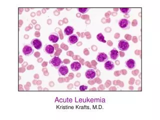
The Hematopoietic & Lymphoid System. Dr. Maha Shomaf. Red Cells Disorders. Types : 1- increase production 2- decrease production. Anemia. Reduction in oxygen transporting capacity of blood mainly due to reduction of the total red cell mass to below normal amounts. Causes of anemia :

E N D
The Hematopoietic & Lymphoid System Dr. Maha Shomaf
Red Cells Disorders • Types : • 1- increase production • 2- decrease production
Anemia • Reduction in oxygen transporting capacity of blood mainly due to reduction of the total red cell mass to below normal amounts .
Causes of anemia : • 1- excessive bleeding • 2- increased RBCs destruction • 3- decreased RBCs production
Anemia due to blood loss • 1- acute : trauma • 2- chronic : GIT lesions & GYN problems
Anemia due to increased destruction (hemolytic anemia) • 1-intrinsic (intracorpuscular) abnormalities). a- hereditary membrane abnormalities enzyme deficiency disorder of Hg synthesis
b- acquired membrane defect (PNH)
2- Extrinsic (extracorpuscular abnormalities) a- antibodies mediated b- mechanical trauma to RBCs e.g -microangiopathic hemolytic anemias: TTP, DIC -intravascular Infections: malaria
Anemia due to impaired production • 1-disturbance of proliferation & differentiation of stem cells aplastic anemia, pure red cell aplasia
2-disturbance of proliferation &maturation of erythroblasts a-defective DNA synthesis: megaloblastic anemias b-defective Hg synthesis Deficient heme synthesis: iron deficiency anemia Deficient globin synthesis: thalassemias: Anemia of renal failure c-unknown or multiple mechanisms
Anemias also can be classified depending on the morphology of the RBCs into : • 1-normocytic • 2-microcytic • 3-macrocytic
Or degree of hemoglobinization into : • 1-Normochromic • 2-Hypochromic • Or RBC shape : • 1-spherocytosis • 2-Ovalocytosis • 3-stomatocytosis • 4-elliptocytosis
Results • 1-Erythroid hyperplasia • 2-Extramedullary hematopoiesis (liver,spleen ,LNs) • 3-Reticulocytosis
RBCs indices are measured by: 1-MCV: the average volume/RBC femtoliters(cubic microns) 2-MCH: the content(mass) of Hg /RBC picogram 3-MCHC:the average concentration of Hg in a given vol. of packed RBCs grams/dl
4- RDW: red cell distribution width the coefficient of variation of RBC volume 5- HCT(hematocrit): the percentage of RBCs in a known volume of blood
Clinical features of anemia • 1-pallor • 2-fatique • 3-lassitude • 4-hyperbilirubinemia & jaundice • 5-GB stones • 6-secondary hemochromatosis • 7-growth retardation • 8-skeletal abnormalities • 9-cachexia
Hemmorhage • If acute it can lead to hypovolemic shock • Hemodilution begins at once & full effect within 2-3 days • The anemia is normocytic normochromic • Recovery is inhanced by ↑ erythropoietin level • The marrow response is marked by reticulocytosis
Chronic blood loss is associated with iron deficiency leading to chronic anemia of under production
Anemia of diminished erythropoiesis
Iron deficiency anemia • Anemia affects 25-50% of the population of developing countries &10% of population of developed countries • IDA is the most common cause of nutritional deficiency anemia
Total body iron is 2gms in women 6gms in men • Distribution : 1- 80% in Hg 2- 20% in myoglobin & iron-containing enzymes (catalase &cytochromes)
Iron stores : -Hemosiderin & Fe-binding ferritin contain 10-20% of total body iron -Stored Fe is mainly found in liver ,spleen & BM -Transferrin is 33% saturated with iron normally -serum iron in men is 120 µg/dL in women 100 µg/dL - (TIBC) total iron binding capacity of serum is 300 -350 µg/dL
There is no regulated pathway for iron excretion • 1-2 mg/d are lost by shedding of mucosal & skin epithelial cells • Iron balance is largely regulated by regulating the absorption of dietary iron • Normal daily western diet contains 10-20mg of Fe mostly heme containing animal products & the remainder as inorganic Fe in vegetables
About 20% of heme Fe is absorbable • Only 1-2% of non-heme Fe is absorbable • The average western diet contains sufficientFe to balance the daily loss loss
Iron absorption • Iron is absorbed in duodenum • Nonheme iron is reduced by ferric reductase then transported by the divalent metal transporter (DMT 1) into the cytoplasm • Iron is transferred to transferrin in the plasma through the action of 2 proteins: • 1-ferroportin • 2-hephaestin
Both DMT 1& ferroportin are widely distributed in the body • Only a fraction of the iron that enters the body is deliverd to transferrin by the action of ferroportin • The remainder is bound to ferritin & lost through the exfoliation of mucosal cells
There is balance between the amount of iron deliverd to transfferin & the iron loss • The balance is regulated by hepcidin synthesized by the liver in an iron- dependent fashion • Hepcidin causes degradation of ferroportin & thus when hepcidin level is high less iron is transferred to transferrin
When hepcidin levels are low iron transport is enhanced as in IDA & ineffective erythropoiesis
Causes of iron deficiency anemia • 1-low dietary intake • 2-malabsorption • 3-increased demands not met by increased uptake • 4-chronic blood loss MENORRHAGIA PEPTIC ULCER STOMACH CANCER ULCERATIVE COLITIS INTESTINAL CANCER HAEMORRHOIDS
Clinical manifestation • Mostly asymtomatic • Weakness • Pallor • Glossitis , stomatitis • Dysphagea • Atrophic gastritis • Dry skin • Hair loss
Thinning & spooning of finger nails (koilonychia) • Pica : the compunction to eat non-foodstuffs such as dirt or clay
Stages of IDA • Iron depletion – Stage One • Iron deficient erthyropoiesis – Stage Two • Iron deficiency anemia – Stage Three
Stage 1 (prelatent) • Iron storage is exhausted - indicated by decrease in serum ferritin levels • Hb (N), MCV (N), iron absorption (), transferrin saturation (N), serum ferritin (),marrow iron () • No anemia – RBC morphology is normal • May have elevated RDW
Stage 2 ( Latent ) • Insufficient iron to insert into protoporphyrin ring to form heme • Protoporphyrin accumulates in cell and complexes with zinc to form ZPP • Hb (N), MCV (N), serum ferritin (), transferrin saturation (), TIBC (), • marrow iron (absent) • No anemia, no hypochromia, but slight microcytosis
Stage 3 • All laboratory tests for iron status become abnormal • Most significant finding is microcytic,hypochromic anemia
Diagnosis • Microcytic hypochromic anemia • ↓ MCV & MCHC • ↓ serum ferritin & iron levels • Low transferrin saturation • ↑ TIBC • ↑ platelets • BM cellularity is only mildly increased although erythropoietin is increased • Extramedullary hematopoiesis is uncommon
Anemia of chronic disease • The most common type of anemia in hospitalized patients • It is related to inflammation-induced sequestration of iron within the cells of reticuloendothelial system
Causes : 1-chronic microbial infection as - osteomyelitis - bacterial endocarditis - lung abscess 2-chronic immune disorder as RA 3-neoplasms as HD , lung & breast carcinoma
Lab findings • Normocytic normochromic anemia • Or microcytic hypochromic anemia • ↑ iron stores • ↑ serum ferritin • ↓ TIBC