Download

1 / 30
300 likes | 421 Vues
This detailed exploration of eyelid anatomy and physiology outlines the function and structure of the eyelids, emphasizing their protective role for the eyes. Key features include the composition of eyelid tissues, the significance of sebaceous and meibomian glands for tear film stability, the mechanics of blinking, and neurovascular supply. Understanding these elements is crucial for ophthalmological practice, surgical interventions, and addressing common eyelid pathologies. This overview serves as a vital resource for students and practitioners in the field of ophthalmology.

E N D
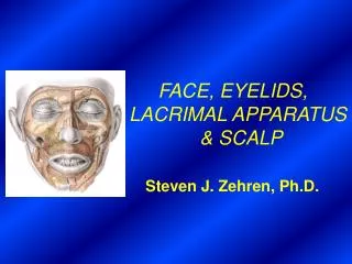
The Eyelids DR. NAILA ALI ASSISTANT PROFESSOR OPHTHALMOLOGY
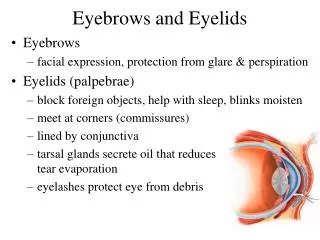
The eyelids are movable folds which act as a shutter protecting eye from injury or excessive light. • Both the upper and lower eyelids meet at medial and lateral canthi with the opening the papebral fissure between them.
SURFACE ANATOMY • Superior PalpebralSulcus • Lateral Canthus • Medial Canthus • Palpebral Fissure
ON THE MEDIAL SIDE • Lacus lacrimalis • Carunculalacrimalis • Plicasemilunaris
The medial 1/6th is distinct and it contains • papilla lacrimalis • punctumlacrimalis • canaliculuslacrimalis.
THE STRUCTURE The margin of each eyelid is 2mm thick and 30mm long It contains eyelashes and just posterior to it are the meibomian glands orifices. The tissues of the lids from anterior (cutancous) to posterior conjunctival aspects as follows:- • skin • subcutaneous areolar tissue • layer of striated muscle (orbicularisoculi) • sub muscular areolar tissue • layer of non striated muscle • the fibrous layer—including tarsal plate • conjunctiva
THE SKIN • It is thinnest and marked by sulci. • The superior sulci is due to aponeurosis of levalor palpebrae superior inserted into the skin. • The less obvious lower sulci is due to the skin being tethered to the underlying periosteum. • Histologically it consist of epidermis, sebaceous and sweat gland and melanocytes.
EYE LASHES • 100 in upper lid • 50 in lower lid • Originate from anterior lamella in two or three irregular rows. • The upper lid lashes are directed upward, and outwards • The lower lid lashed are directed downward and outwards
THE SUBCUTANEOUS AREOLAR TISSUE • It is a loose connective tissue containing no fat. • It is absent at medial and lateral angles, ciliary margin and at sulci.
THE ORIBICULARIS OCULI PartPositionFunction • Orbital Surrounds the orbital Forced lid closure rim • Preseptal In front of the orbital pull lacrimal fascia Septum laterally and create a relative vacuum in lacrimal sac. • Pretarsal in front of the tarsal Close lid and pull Plate lacrimalpuncta medially
THE FIBROUS LAYER---ORBITAL SEPTUM AND TARSAL PLATE • Attached to the orbital margin. • Lies post to the medial palpebral ligament and lateral palpebralraphe medially and laterally. • With in the lids it is thickened to form tarsal plates—embedded in it are tarsal glands.
THE LIGAMENTS • The medial palpebral ligament attaches the medial end of tarsi to lacrimal crest and frontal process of maxilla. • The lateral palpebral ligament attach the lateral end of tarsi to margin tubercle on marginal tubercle of zygomatic bone.
LEVATOR PALPEBRAE SUPERIOR • Originate from lesser wing of sphenoid bone and is inserted an aponeurosis on the ant surface of superior tarsal plate, skin, lat palpebral ligament, medial palpebral ligament • From its inferior surface arises the superior tarsal muscle.
CONJUNCTIVA • Thin mucous membrane lined by non keratinized stratified squamous epithelium. • It has a richly vascularizedSubstantiapropria • At the margin of the eyelids it is continuous with the skin.
ARTERIAL SUPPLY • The lateral palpebral Artery---lacrimal artery. • medial palpebral artery---Ophthalmic Artery. • Marginal and Peripheral arcades.
VENOUS DRANAGE • Medially – Ophthalmic and angular vein • Laterally-superficial temporal vein
NERVE SUPPLY • Upper eyelid • Supra orbital nerve • Supra trochlear nerve • Infra trochlear nerve • Lacrimal nerve • Lower eyelid • Infra trochlear nerve • Infra orbital nerve
SECRETION OF THE EYELIDS Glands Tear Film Layer Location of glands • Meibomian gland Oily layer Tarsal plate • Glands of Zei’s Oily layer Eyelashes • Glands of Wolfring Aqueous layer Tarsal plate • Glands of Krause Aqueous layer Fornix
Blinking There are two main types of blinking • Reflex blinking • Spontaneous Blinking
Reflex Blinking Tactile • Corneal Touch • Cortical Connection • Dimunation of sensitivity in contact lens wearer Dazzle • Bright light • Optic nerve--- Superior colliculus • Associated fiber to facial nuclei Menace • Sudden presence of near object • Optic nerve--- Cortical Connection • Predominantly cortical in nature
Spontaneous Blinking • Occurs at regular basis without an apparent external stimuli. • Mechanics facilitates the drainage of tear film. • Present in blind as no retinal stimuli are required.
Blephrospasm • Simultaneous forcible contraction of orbiculars oculi. • Forcible closure of the lids. • Its role in surgical procedures. • Anterior segment injury.