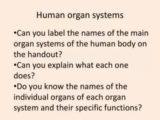
Organ Systems Form meets Function
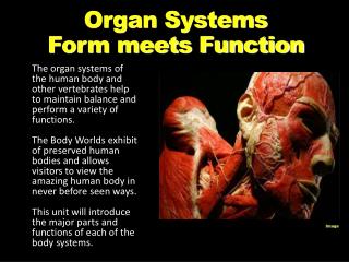
Organ Systems Form meets Function. The organ systems of the human body and other vertebrates help to maintain balance and perform a variety of functions. The Body Worlds exhibit of preserved human bodies and allows visitors to view the amazing human body in never before seen ways.

Organ Systems Form meets Function
E N D
Presentation Transcript
Organ SystemsForm meets Function The organ systems of the human body and other vertebrates help to maintain balance and perform a variety of functions. The Body Worlds exhibit of preserved human bodies and allows visitors to view the amazing human body in never before seen ways. This unit will introduce the major parts and functions of each of the body systems. Image
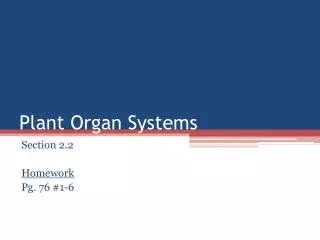
Levels of Organization • The levels of organization in a multicellular organism include cells, tissues, organs, and organ systems • Cellsthe basic unit in living things; specialized cells perform particular functions (EX heart cell) • Tissuesare groups of similar cells that perform a single function (EX connecting muscle to bone) • AnOrganis a group of tissues that work together to perform a complex function (EX Eyes for sight) • An organ systemis a group of organs that perform closely related functions (EX the digestive system) Image
Cells • Cells can be specialized (have a certain function • Function = job • Function is related to the cell structure • Structure = how parts of the cell are put together • Shape • Material it’s made from • Structure of a brain cell is different from muscle cell • Can you tell which cells are neurons, fat, leukocytes, bone (osteocytes, skeletal muscle, smooth muscle, cardiac muscle, cubodial (roll up to make tubes)
Types of Tissues • There are four basic types of tissues in the human body • Epithelial tissue - Glands and tissues that cover interior and exterior body surfaces • Connective tissue - Provides support for the body and connects its parts • Nervous tissue - Transmits nerve impulses throughout the body • Muscle Tissue - Along with bones, helps the body to move
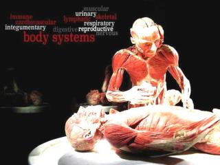
Organ Systems There are 11 organ systems of the human body that work together to maintain homeostasis in the body Homeostasis is the process by which organisms keep internal conditions relatively stable despite changes in external environments Muscular system Skeletal system Nervous system Circulatory system Respiratory system Endocrine system Lymphatic/Immune system Digestive system Excretory system Integumentary system Reproductive system Image
Function: Works with the skeletal system to produce voluntary movement; helps to circulate blood and move food through the digestive system Major Structures: Skeletal Muscles – usually attached to bones and help with voluntary movement Smooth Muscles – found in the walls of hollow structures (stomach, blood vessels, intestines) and NOT under voluntary control Cardiac Muscles – found only in the heart and NOT under voluntary control Works Closely With: the skeletal system to move the body, with the help of signals from the nervous system Muscular System Image
Organization of the Skeletal Muscle Muscle Anatomy If you were to take one whole muscle and cut through it, you would find the muscle is covered in a layer of connective muscle tissue known as the Epimysium that protects the muscle from friction against other muscles and bones.
Organization of the Skeletal Muscle Surrounding the muscle fiber is the Sarcolemma = fibers cell membrane then the Sarcoplasm = cells cytoplasm, containing Glycogen, Fats and Mitochondria for energy. Each muscle fiber itself contains cylindrical organelles known as Myofibrils. Myofibrils made up of bundles of Actin and Myosin proteins which run the length of the muscle fiber and are Important in muscle contraction known as the sliding filament theory.
SKELETAL SYSTEM Hydrostatic Three types of Skeletal systems are: Exoskeleton Endoskeleton
Function: Supports the body; locomotion of voluntary muscles, protection of organs; helps to maintain calcium levels; provides a site for blood cell formation Major Structures: Bones, joints, cartilage, ligaments, tendons Types of Cells: Osteoblasts – build and produce new bone Osteoclasts – break down bone Bone Marrow – within the hollow center of bones, produces red and white blood cells and platelets Works Closely With: the 206 bones in the adult body works with the muscular system to move the body Skeletal System Image
Skeletal System • In the outline of the Homo sapien on your Skeletal Systems page draw and label the following structures: clavicle, femur, fibula, humerus, patella, pelvis, radius, ribs, scapula, skull, sternum, tibia and ulna.
Nervous System Function: Recognizes and coordinates the body’s responses to changes in its internal and external environment Major Structures: Central Nervous System = brain & spinal cord and Peripheral Nervous System = cranial nerves, ganglia and spinal nerves Types of Cells: Neurons – send the messages of the nervous system though electrical impulses Works Closely With: sensory receptors and the five senses (sight, sound, smell, taste, and touch) to interpret stimuli from the environment Image
Central and Peripheral Systems • Central Nervous System (CNS) consists of the brain and spinal cord. • - sensory information goes down the dorsal roots • - motor information goes down the ventral roots to the muscles and glands • dorsal root ganglion (ganglia, plural) • "Gray matter" in middle = cell bodies • "White matter" surrounding = insulated axons
Central and Peripheral Systems • Peripheral Nervous System (PNS) receives signals from the spinal cord and transmits the message by way of peripheral nerves. Peripheral nerves in the cervical region serve the neck and arms; those in the thoracic region serve the trunk; those in the lumbar region serve the legs; and those in the sacral region serve the bowels and bladder. • The PNS consists of • somatic nervous system that connects voluntary skeletal muscles with cells specialized to respond to sensations, such as touch and pain • autonomic nervous system is made of neurons connecting the CNS with internal organs. It is divided into • - sympathetic nervous system which prepares the body for action: fight or flight • -parasympathetic nervous system helps to restore the body, build up energy & supplies needed in the future, and relax
Typical Neuron and Synapse cont • Read the excerpts from the article entitled “Neuron” and very briefly describe the four steps of a nerve impulse down a neuron.
Reflex Arc • A reflex arc is the pathway that a nerve reflex, such as the knee jerk reflex, follows. A tap on the knee stimulates sensory receptors, generating a nerve signal. The signal travels along a nerve to the spinal cord. In the spinal cord, the signal is transmitted from the sensory nerve to a motor nerve. The motor nerve sends the signal back to a muscle in the thigh. The muscle contracts, causing the lower leg to jerk upward. The entire reflex occurs without involving the brain.
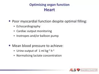
Circulatory System Function - Brings oxygen, nutrients, and hormones to cells; fights infection; removes cells wastes; helps to regulate body temperature Major Structures - Heart, vascular system made up of blood vessels (arteries & veins), blood Heart Video Types of Cells - Red blood cells – transport O2 & CO2 White blood cells – fight infection Platelets – allow blood to clot and stop bleeding Works Closely With: therespiratory system in gas exchange; digestive system to pick up and carry nutrients to the cells of the body the excretory system to filter and clean the blood the endocrine system to deliver hormones Image
Close Up of a Blood Vessel • The connective blood vessels of the body carry the cells of the circulatory system • The vessels can sometimes become blocked with plaque (fatty buildup) shown in yellow Image
Sounds of the Circulatory System • The heart muscle contacts an average of 72 times per minute, sending blood throughout the body through a series of blood vessels. • Sound File Image
Respiratory System Function: Provides oxygen needed for cellular respiration and removes excess carbon dioxide from the body Major Structures: Upper respiratory tract – the nasal cavity, pharynx and larynx Lower respiratory tract – the trachea, bronchi and lungs Key Parts: Nose and nasal cavities, mouth, larynx, trachea, bronchi and their branches, diaphragm, and the alveoli • Works Closely With: thecirculatory system in gas exchange and the muscular system for inhalation and exhalation.
Parts of the Respiratory System • Within each lung, the tiny alveoli are surrounded by blood vessels and oxygen and carbon dioxide diffuse in and out of the vessels. • With each breath, air enters our body through the air passageways and fills up our lungs. Image
Digestive System • Function: • Converts foods into simpler molecules that can be used by the cells of the body; absorbs nutrients; eliminates fecal matter • Major Structures: • Mouth, pharynx, esophagus, stomach, small and large intestines, rectum • Key Parts: • Villi – folded structures within the walls of the intestines which allow for nutrient exchange • Works Closely With: circulatory system to deliver nutrients to the cells of the body Image
Close UP of Digestive Villi • The villi projections allow as much of the nutrients in the digestive system to move in to the circulatory system, providing energy for cells. Image
Digestive Enzymes The pH in the human digestive tract varies greatly. The pH of saliva is usually between 6.5 - 7.5. After we chew and swallow food it enters the stomach, pH 4.0 - 6.5. This is where "predigestion" occurs. Just before leaving the stomach, near the pyloric sphincter, hydrochloric acid (HCI) and pepsin are secreted reaching a pH between 1.5 - 4.0. Food mixes with these juices and enters the small intestine where the pH changes to 7.0 - 8.5. This is where 90% of the nutrients are absorbed and the waste products are passed out through the large intestine, pH 4.0 - 7.0.
Excretory System • Function: • Eliminates urine and other by-products from the body while maintaining homeostasis • Major Structures: • Skin, lungs, kidneys, ureters, urinary bladder, urethra • Key Parts: • Kidneys – remove waste products from the blood • Bladder – collects urine (wastes filtered through the kidney) • Works Closely With: the circulatory system to filter and clean the blood Image
phagocytic leukocyte Fighting theEnemy Within! Immune / LymphaticSystem lymphocytes attacking cancer cell lymph system
Why an immune system? • Attack from outside • lots of organisms want you for lunch! • animals are a tasty nutrient- & vitamin-packed meal • animals must defend themselves against invaders (pathogens) • Viruses, bacteria, Fungi, Protists • Attack from inside • cancers (abnormal body cells) • Function • protects body from disease • collects fluid lost from blood vessels & returns it to the circulatory system Mmmmm, What’s in your lunchbox?
Works closely with the circulatory system to fight infection and collect excess fluids Lymph system Major Structures: lymph vessels (intertwined amongst blood vessels) lymph node
Development of Red & White blood cells Red blood cells inflammatory response fightparasites Leukocytes White blood cells Lymphocytes short-lived phagocytes 60-70% WBC develop into macrophages
Innate vs Acquired Immunity • ACQUIRED • develops only after exposure to microbes, abnormal body cells, toxins or other foreign substances • highly specific because Lymphocytes (white blood cells) produce two types of immune responses • Humoral: cells derived from B cells secrete defensive proteins call antibodies • Cell-mediated: T cells directly destroy infected body and cancer cells, and foreign tissue INNATE • present before any exposure to pathogens • effective from the time of birth • largely nonspecific and slow to respond to specific microbes External: Skin, Mucous membranes, Secretions Internal: Phagocytic Cells, Antimicrobial proteins, Inflammatory response and Natural Killers
Lines of defense • 1st line: Non-specific barriers • broad, external defense • “walls & moats” • skin & mucous membranes • 2nd line: Non-specific patrols • broad, internal defense • “patrolling soldiers” • leukocytes = phagocytic WBC • 3rd line: True immune system • specific, acquired immunity • “elite trained units” • lymphocytes & antibodies • B cells & T cells Bacteria & insectsinherit resistance. Vertebratesacquire immunity.
1st line: Non-specific External defense Lining of trachea: ciliated cells & mucus secreting cells • Barrier • skin • Traps • mucous membranes, cilia,hair, earwax • Elimination • coughing, sneezing, urination, diarrhea • Unfavorable pH • stomach acid, sweat, saliva, urine • Lysosome enzyme • digests bacterial cell walls • tears, sweat
2nd line: Non-specific patrolling cells bacteria • Patrolling cells & proteins • attack pathogens, but don’t “remember” for next time • leukocytes • phagocytic white blood cells • macrophages, neutrophils, natural killer cells • complement system • proteins that destroy cells • inflammatory response • increase in body temp. (fever) • increase capillary permeability • attract macrophages macrophage yeast
Inflammatory response (local non—specific trigger when tissue is damaged)
3rd line: Acquired (active) Immunity • Specific defense with memory • lymphocytes • B cells • T cells • antibodies • immunoglobulins • Responds to… • antigens • cellular name tags • specific pathogens • specific toxins • abnormal body cells (cancer) B cell
Y Y Y Y Y Y Y Y Y Y Y Y Y Y Y Y Y Y Y Y Y Y Y Y Y Y Y Y Y Y Y Y Y Y Y Y Antibodies Y Y • Proteins that bind to a specific antigen • multi-chain proteins • binding region matches molecular shape of antigens • each antibody is unique & specific • tagging “handcuffs” • “this is foreign…gotcha!” Y Y Y antigen-binding site on antibody Y antigen Y Y Y variable binding region Y Y each B cell has ~50,000 antibodies
bone marrow Lymphocytes • B cells • mature in bone marrow • humoral response system • “humors” = body fluids • attack pathogens still circulating in blood & lymph • produce antibodies • Types: Plasma and Memory cells • T cells • mature in thymus • cellular response system • recognize and attack invading cells • Types: Helper, Killer and Memory T Cells
Vaccinations • Immune system exposed to harmless version of pathogen • stimulates B cell system to produce antibodies to pathogen • “active immunity” • rapid response on future exposure • creates immunity without getting disease! • Most successful against viruses
Albert Sabin 1962 oral vaccine 1914 – 1995 Jonas Salk April 12, 1955 • Developed first vaccine • against polio • attacks motor neurons
Polio epidemics 1994: Americas polio free
Passive immunity • Obtaining antibodies from another individual • maternal immunity • antibodies pass from mother to baby across placenta or in mother’s milk • critical role of breastfeeding in infant health • mother is creating antibodies against pathogens baby is being exposed to • Injection • injection of antibodies • short-term immunity (rabies shot)