Download
1 / 58
650 likes | 1.23k Vues
Diabetic foot and peripheral vascular diseases of the foot. Presenter: Dr. J. W. Kinyanjui Facilitator: Dr. V. Kireti 17 th May 2012. Diabetic foot Introduction.

E N D
Diabetic foot and peripheral vascular diseases of the foot Presenter: Dr. J. W. Kinyanjui Facilitator: Dr. V. Kireti 17th May 2012
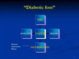
Diabetic footIntroduction • Diabetes mellitus is a group of metabolic diseases characterized by hyperglycemia resulting from defects in insulin secretion, insulin action, or both. • The chronic hyperglycemia of diabetes is associated with long-term damage, dysfunction, and failure of various organs, especially the eyes, kidneys, nerves, heart, and blood vessels. • Diabetic foot is defined as any foot pathology that results directly from diabetes or its long term complications • Two types of diabetes: type I and type II diabetes
Epidemiology • Lesions of the feet affect approx 15% of diabetics in their life with an amputation rate 15 fold higher than non diabetics • Foot ulcerations are the commonest cause of hospital admission in diabetics • Atherosclerosis rarely seen in type I diabetics < 40 yrs while it may be present even before diagnosis in type II • A study conducted at KNH showed that diabetes related gangrene was the indication in 17.5% of lower limb amputations while PVD accounted for 55.3%
Epidemiology – risk factors • Male sex • DM > 10 years duration • Peripheral neuropathy • Abnormal foot structure • Peripheral arterial disease • Smoking • H/O previous ulceration / amputation • Poor glycemic control (HbA1c > 7%)
Pathophysiology • Factors leading to development of diabetic foot: • Diabetic macroangiopathy – peripheral arterial occlusive disease • Diabetic microangiopathy – thickening of basement membranes • Diabetic polyneuropathy • Diabetic osteoathropathy – abnormal foot biomechanics • Reduced resistance to infection • Delayed wound healing • Reduced rate of collateral vessel formation
Diabetic angiopathy • Diabetic macroangiopathy is histologically similar to non diabetic atherosclerosis but distributed in the distal segments of the lower extremities (calf and foot arteries) • Arterial calcification readily detectable on plain x ray with constriction noted on angiography. This compromises oxygen supply to the periphery • Gas exchange is compromised by marked thickening of the capillary basement membrane – a feature of diabetic microangiopathy
Diabetic neuropathy • This affects the sensory, motor, and autonomic fibers • Sensory neuropathy - deep sensory perception is reduced resulting in loss of protective reflexes against physical injury. Typically, manifests in a sock - like distribution. • Motor neuropathy – denervation and atrophy of small foot muscles leading to malum perforans, transverse foot arch instability with clawing and splay foot • Autonomic neuropathy – vasodilation and absent sweating thus foot is warm, dry, scaly which predisposes to fissure formation
Hammer toe Claw toe
Callus formation at pressure points and dry skin are substrate for ulceration
Pes cavus reulting in callus formation over the pressure points
Diabetic neuropathic osteoarthropathy (DNOAP) • Destruction of peripheral and autonomic nerves leads to vasodilation and subsequent demineralization and destabilization of foot skeleton • Sander’s classification based on the location of the lesions in the foot • DNOAP I – necrosis of metatarsophalengeal joints with eventual malum perforans, osteolysis and candystick deformities • DNOAP II – necrosis of the tarsometatarsal joints (Lisfranc’s joint) resulting in a destabilized backfoot. Subluxation of the navicular leads to a clubfoot with abduction of the forefoot and rocking foot deformity. Exposure of the cuneiform-naviculare joint may lead to ulceration at this location
DNOAP I Central malum perforans – MT I and IV Osteolysis MT II Candystick deformity MT III
DNOAP II Ulceration over the navicular-cuneiform joint Destruction of lisfranc articulations Fallen medial arch with navicular subluxation
DNOAP • DNOAP III – necrosis of Chopart’s joint: talo-navicular articulation. Leads to rocking foot deformity where the middle of the sole becomes exposed to pressure. Ulceration occurs directly beneath the verticalized talus. There is as well broadening of the backfoot, abduction of the forefoot and talonavicular subluxation. • DNOAP IV – necrosis of the tibiotalar joint. • DNOAP V – necrosis of the talocalcaneal resulting in a clump backfoot with the bayonet type of deformity.
DNOAP III Ulceration over the navicular Verticalisation of the talus Navicular subluxation Destruction of talonavicular articultaion
DNOAP IV Destruction of tibiotalar joint Lateral malleolar prominence at risk of ulceration
DNOAP V Bayonet deformity Lateral skin ulceration risk Destruction of talo- calcaneal joint
Increased infection rate • Skin fissurations predisposes to penetration of infectious microbes • Polymorphonuclear granulocyte chemotaxis and phagocytosis is impaired • Polyneuropathy predisposes to deep seated infections due to impaired pain sensation • Both anaerobe and aerobe infections are implicated in diabetic foot infections
Examination • Neurological examination • Vibration perception – tuning fork at 128 Hz • Light pressure - Simmes – Weinstein 10 gram monofilament • Light touch • Two point discrimination • Pain • Temperature perception • Deep tendon reflexes • Clonus • Babinski test • Romberg test • Vascular Examination • Palpation of pulses • Skin/limb colour changes • Presence of edema • Temperature gradient • Skin changes • Abnormal wrinkling • Absence of hair • Onychodystrophy • Venous filling time
Examination • Dermatological • Skin appearance • Calluses • Fissures • Nail appearance • Hair growth • Ulceration/infection/ gangrene • Interdigital lesions • Tinea pedis • Markers of diabetes • Musculoskeletal • Biomechanical abnormalities • Structural deformities • Prior amputation • Restricted joint mobility • Tendo Achilles contractures • Gait evaluation • Muscle group strength testing • Plantar pressure assessment
Classification - Wagner • Grade 0 - Skin intact, no foot deformity • Grade 1 - Superficial ulcer • Grade 2 - Deep ulcer • Grade 3 - Deep ulcer with infection • Grade 4 - Limited necrosis • Grade 5 - Necrosis of the entire foot
University of Texas grading • Based on wound ulcer depth and vascular status • Horizontal component: • Stage A – clean wounds • Stage B – non-ischemic infected • Stage C – ischemic non-infected • Stage D – ischemic, infected • Vertical component: • Grade 0 – pre- or postulcerative site that has healed • Grade 1 – superficial wound not involving tendon, capsule or bone • Grade 2 – wound penetrating to tendon or capsule • Grade 3 – wound penetrating bone or joint
Laboratory evaluation • FBS/RBS • Glycosylated hemoglobin (HbA1C) • FHG + ESR • Wound and Blood cultures • Serum Chemistry: CRP • Urinalysis
Imaging Plain X-rays - Osteomyelitis, fractures - Soft tissue gas - Dislocations in neuropathic arthropathy CT Scan Technetium bone scans - osteomyeletis MRI - osteomyelitis
Diabetic foot infection • Divided into uncomplicated non limb threatening infection - superficial cellulitis of limited extension that can be treated on an outpatient basis • Complicated limb threatening infections are more extended and penetrate to deeper tissues, such as tendons, joint capsules, bone or articulations. They require inpatient treatment with surgical debridement and intravenous antibiotics • Osteomyelitis has therapeutic implications such as prolonged antibiotic courses and need for resections
Diabetic foot infection • Superficial swabs overestimate the number of likely microorganisms therefore a deep tissue specimen is preferred as it is more representative • Aerobic gram +ve cocci most common infecting organisms: S. aureus and β-hemolytic streptococci (especially group B) • Chronic wounds have more complex flora: enterococci, enterobactereciae, obligate anaerobes, P. aeuroginosa and other non-fermentative gram negative rods
Management • Preventative foot care • Diabetic foot ulcer (DFU) care • Ischemia management • Neuropathy management • Surgery
Preventative foot care • Podiatry - Regular inspection of the foot, appropriate nail care, warm (32oC) soaks, moisturizing creams, early detection of new lesions • Optimally fitted footwear – well cushioned sneakers, custom molded shoes • Pressure reduction – cushioned insoles, custom orthoses • Patient education — need for daily inspection and necessity for early intervention, avoidance of barefoot walking • Physician education — significance of foot lesions, importance of regular foot examination, and current concepts of diabetic foot management
DFU care • Debridement – of callus and necrotic tissue using sharp debridement till bleeding tissue, lavage and dressings • Offloading of the ulcer site to reduce ischaemia via total contact cast, non weight bearing (crutches, bedrest, wheel chair) • Wound management – maintenance of a moist wound with regular cleaning and dressing • Infections treated with broad spectrum antibiotics based on culture results. Clindamycin/flouroquinolone/metronidazole suitable empiric therapy
Ischemia/neuropathy • Angiography evaluates for chance of catheter intervention or vascular surgery • Vascular bypass surgery successful if occlusion is supramalleolar but less so in inframalleolar PAOD • Aspirin is useful for primary and secondary prevention • Neuropathy treated pharmacologically with agents such as carbamazepine, gabapentin and pregabalin and prevention of minor trauma that will go undetected due to insensate foot
Surgery • Sharp debridement • Local procedures to remove areas of chronically elevated pressure (deformities) causing non healing ulcers • Sequestrectomies • Amputation • Correct structural deformities — hammer toes, bunions, Charcot
Indications for amputation • Uncontrollable infection or sepsis • Inability to obtain a plantar grade, dry foot that can tolerate weight bearing • Non ambulatory patient
Other peripheral vascular diseases • Peripheral arterial occlusive disease (PAOD) • Post thrombotic syndrome • Chronic venous insufficiency
PAOD • Most common cause is atheroclerosis which narrows the lumen of peripheral arteries • Buerger’s disease is a potentially preventable cause due to its association with smoking • Symptoms include: • Intermittent claudication • Ischaemic rest pain • Signs include: • Calf muscles atrophy • Loss of hair growth over the dorsum of the toes • Thickening of the toenails • Atrophy of the skin • Delayed capillary refill • Ischaemic ulcers
PAOD • Ischemic ulcers are painful with a ‘punched out’ appearance. • They are commonly located distally over the dorsum of the foot or toes. • The ulcer base usually consists of poorly developed, grayish granulation tissue. • Critical limb ischaemia is defined as persistent ischemic rest pain lasting for more than 2 weeks and/or ulceration of the leg, associated with an ankle systolic pressure < 50 mm Hg and/or a toe systolic pressure of <30 mm Hg and or an ABPI < 0.9.
PAOD - Investigations • Ankle systolic pressure measurement – 12cm cuff used and doppler probe over the dorsalis pedis or posterior tibial artery. <50mmHg implies critical limb ischemia and aggressive revascularisation needed • Toe systolic pressure – 25mm cuff over proximal phalanx of hallux. Critical limb ischemia at < 30mmHg • Transcutaneous oxygen pressure – electronic probe used. Normal range 30 – 50 mmHg. < 30 mmHg implies critical ischaemia • Doppler ultrasound – operator dependent. More accurate for assessment of femoropopliteal vessels than tibioperoneal arteries • Arteriography – gold standard, invasive, contrast used. Useful where vascular procedures are being planned
Management of PAOD • Secondary prevention – statins, aspirin, DM and HTN control, smoking cessation • Walking excersises 1h/day– reduced intermittent claudication by encouraging collateral vessel formation • Footwear fitted to reduce pressure and increased warmth. May need to be customised. Minimise exposure to cold and mositure • Surgical debridement of ulcers with appropriate dressing thereafter, infection control • Interventional vascular procedures such as percutaneous transluminal angioplasty, bypass procedures indicated in critical limb ischaemia • Amputation may eventually be necessary
Post thrombotic syndrome • Symptoms and signs that typically follow DVT • Caused by post thrombotic recanalisation and valve destruction that leads to chronic ambulatory venous hypertension • Not limb threatening but adversely affects quality of life • Symptoms include chronic leg discomfort • Signs include: • Edema • skin changes – pigmentation, dermatitis, liposclerosis • Ulcers – typically supramalleolar medial, painless, irregular edges
Management • Primary prevention by preventing DVT in at risk patients • Early management of DVT and continued antithrombotic therapy to prevent recurrence of DVT • Fibrinolysis and thrombectomy – controversial due to minimal benefit and increased risk • Compression therapy – effective primary prevention of DVT and secondary prevention of PTS after DVT. Layered compression stockings now the mainstay of treatment • Vascular surgery – not as useful as in varicose veins because PTS is a disease of the deep system
Chronic venous insufficiency • Chronic venous insufficiency (CVI) affects 10-15% of men and 20-25% of women • Characterised by chronic inadequate drainage of venous blood and venous hypertension, which results in • leg edema (swelling) • dermatosclerosis (hardening of the skin) • Feelings of pain, fatigue and tenseness in the lower extremities • May be complicated by • skin ulceration • chronic (and potentially life-threatening) infections of the lower extremities