Quail Induced Pluripotent Stem Cells Derived Using Human Reprogramming Factors
10 likes | 153 Vues
Quail Induced Pluripotent Stem Cells Derived Using Human Reprogramming Factors. Yangqing Lu 1,2,4 , Franklin D. West 1,2 , Brian J. Jordan 3 , Jennifer L. Mumaw 1,2 , Erin T. Jordan 1,2 , Amalia Gallegos-Cardenas 1,2 , Robert B. Beckstead 2 , Steven L. Stice 1,2

Quail Induced Pluripotent Stem Cells Derived Using Human Reprogramming Factors
E N D
Presentation Transcript
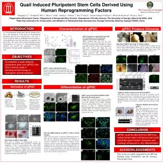
Quail Induced Pluripotent Stem Cells Derived Using Human Reprogramming Factors Yangqing Lu1,2,4, Franklin D. West1,2, Brian J. Jordan3, Jennifer L. Mumaw1,2, Erin T. Jordan1,2, Amalia Gallegos-Cardenas1,2, Robert B. Beckstead2, Steven L. Stice1,2 1 Regenerative Bioscience Center, 2Department of Animal and Dairy Science, 3Department of Poultry Science, The University of Georgia, Athens GA 30602, USA 4State Key Laboratory for Conservation and Utilization of Subtropical Agro-bioresources, Guangxi University, Nanning, Guangxi 530004, China INTRODUCTION Characterization of qiPSC qiPSC Produce Chimeras As a developmental model quail-chicken chimeras have led to the elucidation of key facets in developmental patterning and cell fate, partially due to the ease of access to and the ability to manipulate the avian embryo. Avian pluripotent stem cells and derived committed cell lines offer a cell source which could recapitulate normal development in vitro and in vivo when transplanted into embryos and provide the opportunity to altered development through genetic manipulation of the stem cells. qiPSCs demonstrate rapid proliferation, high levels of telomerase activity and clonal expansion after genetic manipulation qiPSC doubling time was 16.6 hr (n=3), significantly faster than the QEF cells (36.9 hr; P< 0.01) (A). Telomerase activity in qiPSC was higher than QEF (>11 fold, *P<0.01) and comparable to HeLa cells (P=0.07) (B). Eight days post transduction a subpopulation of qiPSCs expressed GFP (C, D, E). Nine days after FACS sorting, clonally expanded GFP+ qiPSCs generated colonies (F, G) that maintained GFP expression long term (H). Dapi AF488:Oct4 Overlay Injection of qiPSC into stage X chicken embryo To inject qiPSCs into chicken embryos, a single window was drilled into the shell of stage X White Leghorn chicken egg (A). qiPSC were then injected into the subgerminal cavity with a micropipette (B). Windows were sealed with hot glue and injected eggs were then transferred to incubators (C). OBJECTIVES To establish a quail induced pluripotent stem cell (qiPSC) line that could be used in developmental biology or transgenic animal research. qiPSCs express pluripotent genes Immunocytochemistry demonstrated that QEFs were negative for POU5F1 (A) and SOX2 (B), while qiPSCs were POU5F1 (D) and SOX2 (E) positive. Scale bars are 50 μm. RESULTS Derivation of qiPSC Differentiation of qiPSC Chimeric chicken embryos derived from qiPSC GFP+ qiPSCs were incorporated into the brain (A, ectoderm), eye (B, ectoderm), trachea/lung (C, endoderm), heart (D, mesoderm) and yolksac (F, extraembryonic tissue) of quail-chicken chimeric embryos. PCR results demonstrated that various tissues and qiPSCs were positive for the hPOU5F1 transgene, but negative in chicken embryonic fibroblast (CEF) (E, G). qiPSCs generate Embryoid Bodies (EBs) that form all 3 germ layers Compact EBs were formed after 6 days in culture (A). EBs were replated for further differentiation for 2 days (B). Ectoderm (TUJ1, PAX6), endoderm (Vimentin) and mesoderm (Brachyury) genes were expressed in EBs (C). Immunocytochemistry demonstrated that EB derived cells were positive for ectoderm (TUJ1, D), endoderm (SOX17, E) and mesoderm (αSMA, F) proteins (Dapi merge D’-F’). Scale bars are 50 μm. QEFs underwent lentiviral transduction utilizing viPS kit (Thermo Scientific) containing the human stem cell genes POU5F1, NANOG, SOX2, LIN28, KLF4 and C-MYC. qiPSCs were observed around 17 days after transduction growing as compact colonies. qiPSCs were manually harvested and plated on Matrigel coated dishes in mTeSR1 medium Quail embryonic fibroblast cells (QEFs) were isolated from day 11 embryos and cultured in DMEM. Chimeric chickens derived from qiPSCs Two chicks produced by low passage qiPSC (P7) determined to be at day 14 (A) and 19 (B) developmental stages exhibited significant levels of feather chimerism (black arrow), yet failed to hatch. High passage qiPSC (P45) contributed to live chimeras as indicated by the presence of the human POU5F1 gene in the brain, liver and gonad of two individuals out of 15 examined individuals ( C). CONCLUSION Directed differentiation of qiPSCs to three neural lineages. qiPSCs were subjected to a 3 step neural differentiation process, with cells first cultured in neural derivation medium for 12 days, then in proliferation medium for 7 days, followed by continual maintenance in differentiation medium. Neurite extensions could be found after culturing in differentiation medium for 48 days. Neuron-like cells expressing Hu C/D+ (A) and MAP2 (B) were present after 10 days of differentiation and astrocyte (C) and oligodendrocyte- (D) like cells after 23 and 39 days of differentiation, respectively. Scale bars in E, F and G are 50 μm. Scale bars in D are 100 μm. qiPSC could be derived from QEFs by using human stem cell factors and could generate quail-chicken chimeras and undergo advanced in vitro differentiation. Derivation of qiPSCs from QEFs QEFs prior to addition of reprogramming factors (A). Incomplete reprogrammed QEFs maintained a fibroblast-like morphology at day 6 post-transduction (B), while qiPSC colonies at day 17 showed defined borders (C) and at the single cell level, a high nuclear to cytoplasm ratio, clear cell borders and prominent nucleoli (D, E). qiPSCs were positive for AP (F) and PAS (G). 5 out of 6 human pluripotent stem cell factors were integrated and expressed in qiPSCs (H). ACKNOWLEDGEMENTS This work was jointly supported by the Bill and Melinda Gates Foundation and the Guangxi Scholarship Fund.