Download
1 / 17
170 likes | 273 Vues
Learn about the diagnostic criteria for long QT syndrome, causes of QT prolongation, and management strategies including differential diagnoses and treatment options such as defibrillation and temporary pacing.

E N D
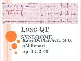
Long QT and TdP Morning Report Elias Hanna, LSU Cardiology
Markedly prolonged QT (QTc~700 ms) • Wide and ample T waves in V2-V4, deep T wave inversion in the inferior leads • T wave alternans
Arrows of different colors point to the T wave of alternating morphology every other beat, called T wave alternans. T wave alternans may be seen in a subgroup of patients with prolonged QT and implies a very heterogeneous repolarization and a more imminent risk of TdP. More typically seen in congenital long QT syndrome
How to we define long QT? • QT> ½ RR • Corrected QT (QTc)>450 ms in M, 460 ms in F
How to calculate corrected QT? • Corrected QT means QT corrected for heart rate. QT normally decreases at faster heart rate. Thus, while QT is normally <460 ms at a rate of 60 bpm, a lower cutoff should be used for a rate of 100 bpm • How to calculate corrected QT: 1-QTc= QT/√RR ( RR in sec, not msec!!) e.g: QT of 400 msec at a heart rate of 100 bpm -At a rate of 100 bpm, RR interval is 600 msec=0.6 sec QTc=400/√0.6=515 msec
2-Another quick method: QTc is the patient’s QT had the heart rate been 60 bpm. Normally, QT 20 ms for every 10 beat in rate, and 20 ms for every 10 beat in rate to calculate QTc, i.e QT at the rate of 60 for this pt, add 20 ms for every 10 beats above 60 if QT is 400 ms at a rate of 100, then QTc= 400+ (4x20)=480 ms (the 2 methods may yield slightly different results)
Differential dx of long QT • Electrolytes (K, Mg, Ca) (+other: hypothyroidism) • Drugs (antiarrhythmics class I, III; macrolide or quinolone antibiotics; antipsychotics…) • Ischemia • Congenital long QT syndrome (LQT 1,2,3)
In this case • The pt had low K (3.0) and Mg (0.7). • However, the shape of ST segment and T wave, particularly the fact that T wave is wide and ample T, does not fit with hypokalemia or hypocalcemia (see next slide for electrolyte shapes) • In light of the ST/T shape, K and Mg abnormalities are not the sole cause of QT prolongation but rather an exacerbating factor • The pt likely has ischemia or congenital long QT syndrome as underlying etiology. In this case, coronary angio did not show any CAD, and QT interval strikingly but remained prolonged (480 msec) after correction of lytesabnormlities congenital long QT syndrome is the underlying cause of his QT prolongation
Typical ST/T shape in hypokalemia: ST depression with prominent T (actually U) and prolonged QT when K<2.5-3 Flat T with K~3
Hypocalcemia: Long QT that is due to a long ST segment, which is different from long QT due to congenital long QT syndrome, drugs, or hypokalemia. T wave is not wide, there is no T wave abnormality.
The ST/T shape in this case is not typical of electrolyte abnormalities, but QT prolongation is certainly exacerbated by these electrolyte abnormalities
Pt goes into this rhythm Torsades de pointe (polymorphic VT with changing QRS polarity, with a long baseline QTc) sinus VF (disorganized and chaotic rhythm, QRS almost vanishes every now and then) Defibrillation
Polymorphic VT vs. TdP • Polymorphic VT with changing polarity of the QRS complexes is not necessarily torsades de point (TdP). • In order to say TdP, you need to have: (1)polymorphic VT as in the prior slide (2) long baseline QT on the ECG obtained before or after TdP • If you have TdP but normal QT, then the rhythm is called polymorphic VT not TdP • The tx of TdP is different from polymorphic VT
Polymorphic VT is usually an ischemic rhythm and is treated with shock, emergent cath/PCI, and amiodarone
TdP is given 3 therapies: 1-Defibrillation 2-Magnesium 2 g IV (regardless of Mg level) + start correcting K 3-Temporary pacing after the run of TdP has resolved. Temporary pacing prevents TdP from recurring: Usually, TdP occurs in a pt with prolonged QT who is also bradycardic. Bradycardia further prolongs QT and furthers disperses repolarization delays across the myocardium. Bradycardia is a major trigger of TdP, particularly TdP in patients with acquired long QT. Pacing to a rate of 80-100 bpm will prevent TdP recurrence. Pacing does not apply to our pt here because he is tachycardic. Congenital long QT, as opposed to acquired long QT, is often triggered by catecholamine surge and may be associated with tachycardia No Amiodarone!!! Amiodarone prolongs QT