Download
1 / 41
410 likes | 425 Vues
Learn about the structure of the heart, blood vessels, and the path of blood flow through the circulatory system. Understand the cardiac cycle and the conduction system of the heart.

E N D
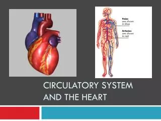
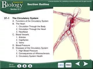
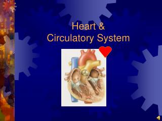
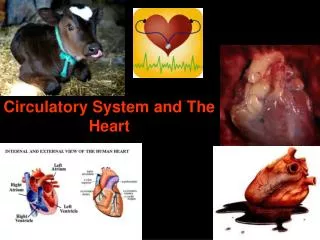
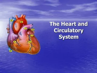
The circulatory system… The cardiovascular system delivers oxygen and nutrients to tissues and removes wastes.
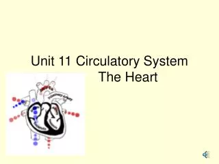
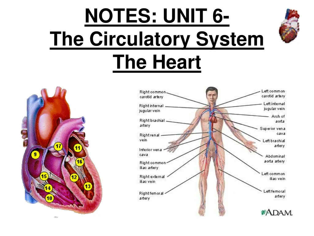
STRUCTURE OF THE HEART Size: ● about 14 cm long and 9 cm wide Location: ● 2/3 left of midline ● below 2nd rib and rests on diaphragm • Location allows for CPR ● divided into right and left sides • Divided by interventricular septum
STRUCTURE OF THE HEART Heart Covering:the heart is enclosed in a double-walled sac called the pericardium (protects against friction) ● loosely fitting superficial part of the sac: fibrous pericardium ● deep to the fibrous pericardium: serous pericardium *composed of 2 layers *space between the layers = pericardial cavity (cushions and lubricates heart)
STRUCTURE OF THE HEART Heart Wall ● epicardium = outermost layer; in contact with pericardium (older people= infiltrated with FAT ) ● myocardium = middle layer; thick, muscular (mainly cardiac muscle tissue!) ● endocardium = inner layer; lines the heart chambers
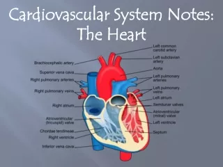
each side is divided into an atrium and a ventricle (blood always flows from atrium to ventricle) • ATRIA: chambers that receive blood returning to the heart • VENTRICLES: chambers that pump blood out of the heart • one-way valves to prevent “backflow” as blood moves through the heart • R and L AV valves (tricuspid/bicuspid) • Pulmonary & aortic semilunar valves
Left AV valve (bicuspid) Right AV valve
● Structure of AV valves: -CHORDAE TENDINAE: strong fibrous structures (“heart strings”) that attach to the flaps of the valves hold AV valves closed while ventricles contract -PAPILLARY MUSCLES: muscles embedded in the endocardium; attach to the chordae tendineae contract to hold valves closed *serve to anchor flaps in their closed position so pressure doesn’t blow them into atria like an umbrella on a windy day
Blood Vessels & Blood Flow ● RIGHT SIDE: • RIGHT ATRIUM receives deoxygenated blood from the superior and inferior vena cava and coronary veins (sinus) • pumps blood into the RIGHT VENTRICLE right ventricle pumps blood out of the heart into the PULMONARY ARTERIES (takes blood to the lungs to get O2)
● LEFT SIDE: LEFT ATRIUM receives oxygenated blood from the pulmonary veins (coming from the lungs) pumps blood into the LEFT VENTRICLE left ventricle pumps blood out of the heart into the AORTA (takes blood to all of the body)
SEMILUNAR VALVES: prevent backflow from arteries leaving the heart -PULMONARY VALVE: separates pulmonary arteries from R ventricle -AORTIC VALVE: separates aorta from L ventricle *ventricle contraction forces these valves open until contraction is over; blood flowing backwards closes valves
2 Types of Circulation ● pulmonary circulation:to the lungs & back blood leaves from right side of heart
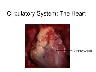
2 Types of Circulation: ● systemic circulation: out to the body & back blood leaves from the left side of the heart *coronary circulation: supplies the heart muscle (myocardium) shortest systemic circuit: blood branches off aorta to coronary arteries, supplies oxygen to myocardium, and then returns through coronary veins & coronary sinus to Right atrium
**500,000 Americans die per year of coronary artery disease **3.5 million Americans have coronary problems
Path of Blood Flow Through the Heart • Right side • Deoxygenated blood enters the right atrium through the superior and inferior vena cava through right atrioventricular (tricuspid) valve right ventricle through pulmonary semilunar valve pulmonary artery arterioles capillaries of lungs • Left Side • Oxygenated blood returns from the capillaries of the lungs through venules pulmonary veins left atrium left atrioventricular (bicuspid, mitral) valve left ventricle aortic semilunar valve aorta
Cardiac Cycle: *systole: contract *diastole: relax ●When the whole heart is in diastole, in Ventricular filling: blood returning to heart passively & flows into atria & through the open AV valves ● ATRIAL SYSTOLE: atria contract (propels residual blood into ventricles) while the ventricles relax (VENTRICULAR DIASTOLE) ● VENTRICULAR SYSTOLE: ventricles contract while the atria relax (ATRIAL DIASTOLE); blood leaves heart ● all chambers relax for a brief period; cycle repeats!
Heart Poster- start • Label the anatomy of the heart • Then color code blood flow using arrows through the heart • BLUE: deoxygenated blood • RED: oxygenated blood
Conduction System of the Heart • The conduction system of the heart is composed of 4 structures • Sinoatrial Node (SA Node): initiates contraction; PACEMAKER • Electrical message passes throughout R & L atria atrial systole • Atrioventricular Node (AV node): receives message from SA Node & slows it down for 1/10 second before passing it through AV bundle (bundle of His)
Conduction System of the Heart • Atrioventricular (AV) Bundle: passes electrical impulse to Purkinje fibers on left and right sides of ventricles • Purkinje Fibers: stimulates the right & left ventricles to contract together ventricular systole
ELECTROCARDIOGRAM (ECG/EKG): ● records the electrical changes in the myocardium during a cardiac cycle ● the pattern has several characteristic waves: 1) P wave: depolarization wave from SA through atria 2) QRS complex: ventricular depolarization; precedes ventricle contraction 3) T wave: ventricular repolarization
A 60 year old woman with 3 hours of chest pain. Acute Myocardial Infarction
A 55 year old man with 4 hours of "crushing" chest pain. Acute Myocardial Infarction
Regulation of Cardiac Cycle: ● Heartbeat is affected by: -Physical exercise -Body temperature -Concentration of ions (calcium, potassium) -Emotions
Heart Poster • Color code blood flow using arrows through the heart • BLUE: deoxygenated blood • RED: oxygenated blood • Label heart Anatomy • Label the 4 structures in the conduction system on your heart • Use yellow highlighter to outline the flow of impulse conduction through the heart