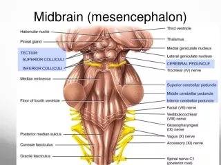
MIDBRAIN
MIDBRAIN. forms a transition (and fiber conduit) to the cerebrum also contains a number of important cell groups, including several cranial nerve nuclei.

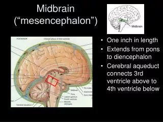
MIDBRAIN
E N D
Presentation Transcript
forms a transition (and fiber conduit) to the cerebrum • also contains a number of important cell groups, including several cranial nerve nuclei.
The base of the midbrain contains the cruscerebri, a massive fiber bundle that includes corticospinal, corticobulbar, and corticopontine pathways • The base also contains the substantianigra
substantianigra • Its cells contain neuromelanin and receives afferent fibers from the cerebral cortex and the striatum • it sends dopaminergic efferent fibers to the striatum • The substantianigra plays a key role in motor control. Degeneration of the substantianigra occurs in Parkinson's disease
The external aspect of the basis of the midbrain is called the cerebral peduncle.
The corticobulbar fibers • from the motor cortex to interneurons of the efferent nuclei of cranial nerves • are homologous with the corticospinal fibers. • fibers to the lower portion of the facial nucleus and the hypoglossal nucleus are crossed (from the opposite cerebral cortex). • All other corticobulbar projections are bilaterally crossed (from both cortices).
The fibers of the oculomotor (III) nerve exit between the cerebral peduncles in the interpeduncular fossa. • The fibers of the trochlear (IV) nerve exit on the other side of the midbrain, the tegmentum
Tegmentum • contains all the ascending tracts from the spinal cord or lower brain stem and many of the descending systems. • A large red nucleus receives crossed efferent fibers from the cerebellum and sends fibers to the thalamus and the contralateral spinal cord via the rubrospinal tract. • The red nucleus is an important component of motor coordination.
Two contiguous somatic efferent nuclear groups lie in the upper tegmentum • the trochlear nucleus (which forms contralateral nerve IV) • the oculomotor nuclei (which have efferent fibers in nerve III).
Tectum [‘roof’] • formed by two pairs of colliculi • The superior colliculi contain neurons that receive visual as well as other input and serve ocular reflexes • the inferior colliculi are involved in auditory reflexes and in determining the side on which a sound originates.
The inferior colliculi receive input from both ears, and they project to the medial geniculate nucleus of the thalamus by way of the inferiorbrachium. • The superior brachium links the lateral geniculate nucleus and the superior colliculus.
The colliculi contribute to the formation of the crossed tectospinal tracts • These are involved in blinking and head-turning reflexes after sudden sounds or visual images.
Periaqueductal Gray Matter • Contains descending autonomic tracts as well as endorphin-producing cells that suppress pain. • This region has been used as the target for brain-stimulating implants in patients with chronic pain.
Superior Cerebellar Peduncle • Contains efferent fibers from the dentate nucleus of the cerebellum to the opposite red nucleus (the dentatorubrothalamic system) and the ventral spinocerebellar tracts. • The cerebellar fibers decussate just below the red nuclei.
Weber's syndrome • in the basal midbrain, involves nerve III and portions of the cerebral peduncle • There is a nerve III palsy on the side of the lesion and a contralateralhemiparesis (because the lesion is above the pyramidal decussation). • The arterial supply is by the posterior perforators and branches of the posterior cerebral artery
Benedikt's syndrome • situated in the tegmentum of the midbrain • may damage the medial lemniscus, the red nucleus, and nerve III and its nucleus and associated tracts • This area is supplied by perforators and branches of circumferential arteries.