Download

1 / 12
120 likes | 252 Vues
Excretory System Bobby Singh & Chudi Mbanefo. The Kidney. 1. Function. The main function of the kidney is to maintain homeostasis of the blood removes toxins created by metabolism or ingested in diet removes excess water

E N D
Excretory System Bobby Singh & Chudi Mbanefo
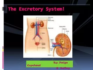
The Kidney 1. Function • The main function of the kidney is to maintain homeostasis of the blood • removes toxins created by metabolism or ingested in diet • removes excess water • Renal Pelvis sends all produced waste (urine) to the ureter to be released • Urine flows down the ureter to the bladder where it is stored until release • When bladder is ready to release urine, the urethra opens and allows the bladder to empty
The Kidney (cont.) Structure • Every human has TWO kidneys made up of millions of Nephrons. • Renal Artery carries in blood with waste • Renal Vein carries out blood without waste • There are three parts in a kidney: • renal cortex • renal medulla • renal pelvis • RENAL: pertaining to the kidney • Major calyces are found all throughout the kidney and carry all collected waste
The Renal Structures • Renal Cortex: • Outer layer of kidney, its extensions, renal columns, extend into the renal medulla • part of the kidneys made of nephrons and blood vessels. • main function is to purify the blood and clear the body of waste products. • located between the capsule and the medulla and can be recognized by its pale color. • Renal Medulla: • Located deep to the cortex and • The inner most portion of a kidney • Consists of multiple renal pyramids (cone shaped) • Tips of the cones (papillae) project into the minor calyces of the renal pelvis.
The Renal Structures (cont.) • Blood vessels from the renal artery are found on the border of the medulla and provide circulation for the kidney • blood passes through the medulla, the last stop for blood going through the kidney 3. Renal Pelvis: • where flow of the urine begins • connects the main body kidney to the ureter • place where all the major calyces meet up to get down
The Nephron Basic functional unit of the kidney • made up of a knot of coiled capillaries • Glomerulus • Small blood vessel that looks like a ball of yarn. • The filtering actually occurs in the glomerulus. • Acts as a filter, keeping in normal proteins and cells and letting out wastes. • Tubule • Also called the renal or kidney tubule, is the tube where the wastes filtered out from the glomerulus pass through Function • Removes extra, unnecessary water, wastes and other substances from your blood. • Replenishes substances like sodium, potassium or phosphorus whenever any of these run low in your body.
The Ureter Ureter Structure: • tubes made of smooth muscle • when fully developed, ureters are usually 10–12in long (diameter of ureters are half the diameter of an average capillary!) Ureter Function: • pump urine from the kidneys to the urinary bladder • connects to kidney by way of the renal pelvis • In males, the ureter is found on genital-fold while in females it is found on the uterosacral ligament.
The Bladder Breaking Down the Urge • large sack of smooth muscle that holds urine until the body is ready, or forced, to release it • stretch receptors in the sack create the “I GOTTA PEE NOW,” feeling • When emptied the bladder is about the size and shape of a pear, the walls collapse into folds called rugae • the average human bladder can hold 400-600 mL Disorders/Conditions • Cystitis: also called Painful Bladder Syndrome (PBS) inflammation in the bladder causing chronic pain • Urinary Retention: Urine does not exit the bladder consistently due to issues with muscle in the bladder. The bladder may swell to hold more than a quart of urine. • Cytocele: (women only) Weakened pelvic muscles, usually from childbirth, allow the bladder to press on the vagina. Creates complications with urination. • Bedwetting: chronic urination while sleeping common in children up to 5 years old. In older cases it is considered “being a little baby.” Nothing to be ashamed of though.
In females, the urethra is 1.5 inches, but in males, they reach out to be 8 INCHES! The Urethra • Release path from the bladder for the created urine • Has two sphincters to regulate the flow of urine by opening and closing • Internal: involuntary/no control, smooth muscle that is the same in both males and females • External: voluntary/some control, skeletal muscle that differs by gender • females- made up of three muscles that work together that constrict together to squeeze the vagina and the urethra at the same time • males- functions the same as the internal sphincter
Kidney Diseases • Kidney Failure • takes place when kidneys begin to not function properly • Results in accumulation of waste products and toxic products • Kidney begins to function less than 20% • May cause permanent damage to body cells, tissues, and organs • Kidney Stones • Small hard deposits forming inside the kidney • Usually form when urine becomes concentrated, allowing minerals to crystallize and stick together. • No permanent damage is caused and a simple way to rid the stone is by drinking lots of water and taking pain medications. • May need surgery in certain cases.
Kidney Diseases • Diabetic Kidney Problems • Diabetes causes your blood sugars to be too high • Kidneys are unable to properly filter the rising amounts of sugar • One of the main causes of kidneys failure • Urinary Tract Infection (UTI) • Can occur in any part of your urinary system (kidneys, ureters, bladder and urethra) • Caused by germs getting into the urinary tract system through the urethra • Women are at greater risk to develop UTI than men because of their shorter urethra • Antibiotics are the basic treatment for these infections • If not treated quick enough, it can damage your kidneys and lead to a kidney infection as well
Works Cited • "The Bladder (Human Anatomy): Function, Picture, Location, Definition." WebMD. WebMD, 1 July 2009. Web. 11 May 2013. <http://www.webmd.com/urinary-incontinence-oab/picture-of-the-bladder>. • "Excretory System." ScienceDaily. ScienceDaily, n.d. Web. 11 May 2013. <http://www.sciencedaily.com/articles/e/excretory_system.htm>. • Mangusan, David, Jr. "Kidney Health Care (KHC)." The Nephron. N.p., n.d. Web. 11 May 2013. <http://www.kidneyhealthcare.com/2010/12/nephron-structure-function-nephron.html>. • Riebe, Mary. "The Structure of the Kidney." The Structure of the Kidney. N.p., n.d. Web. 12 May 2013. <http://www.wisc- online.com/objects/ViewObject.aspx?ID=NUR2503>. • Spence, Alexander P. Basic Human Anatomy. Redwood City, CA: Benjamin/Cummings Pub., 1982. Print. • Walker, Richard. Firefly Guide to the Human Body. Toronto: Firefly, 2004. Print. • "What Is the Renal Cortex?" WiseGEEK. WiseGEEK, n.d. Web. 11 May 2013. <http://www.wisegeek.org/what-is-the-renal- cortex.htm>. • YouTube. Perf. Khan Academy. YouTube. YouTube, 03 Mar. 2010. Web. 12 May 2013. <http://www.youtube.com/watch?v=cc8sUv2SuaY>.