CH 12 NOTES, part 1 : Chromosomes, the Cell Cycle, and Cell Division (12.1-12.2)
560 likes | 1.28k Vues
CH 12 NOTES, part 1 : Chromosomes, the Cell Cycle, and Cell Division (12.1-12.2). ● The ability of organisms to reproduce best distinguishes living things from nonliving matter ● The continuity of life is based upon the reproduction of cells, or cell division.

CH 12 NOTES, part 1 : Chromosomes, the Cell Cycle, and Cell Division (12.1-12.2)
E N D
Presentation Transcript
CH 12 NOTES, part 1: Chromosomes, the Cell Cycle, and Cell Division (12.1-12.2)
● The ability of organisms to reproduce best distinguishes living things from nonliving matter ● The continuity of life is based upon the reproduction of cells, or cell division
● In unicellular organisms, division of one cell reproduces the entire organism ● Multicellular organisms depend on cell division for: Development from a fertilized cell Growth Repair
100 µm 200 µm 20 µm Reproduction Growth and development Tissue renewal
12.1 – Most cell division results in genetically identical daughter cells ● Cells duplicate their genetic material (DNA) before they divide, ensuring that each daughter cell receives an exact copy of the genetic material ● A dividing cell duplicates its DNA, allocates the two copies to opposite ends of the cell, and only then splits into DAUGHTER CELLS
Cellular Organization of the Genetic Material: ● A cell’s endowment of DNA (its genetic information) is called its GENOME ● DNA molecules in a cell are packaged into CHROMOSOMES
● Every eukaryotic species has a characteristic number of chromosomes in each cell nucleus ●Somatic (nonreproductive) cells have two sets of chromosomes (DIPLOID) ●Gametes (reproductive cells: sperm and eggs) have half as many chromosomes as somatic cells (HAPLOID) ● Eukaryotic chromosomes consist of CHROMATIN, a complex of DNA and protein (i.e. histone proteins) that condenses during cell division
● Chromosomes = after the DNA replicates in the S phase of interphase, a chromosome consists of tightly coiled chromatin (DNA); ● a chromosome consists of 2 identical chromatids(sister chromatids) which are connected in the center by a CENTROMERE **a human cell entering mitosis contains 46 chromosomes
chromosome centromere chromatin DNA
0.5 µm Chromosome duplication (including DNA synthesis) Centromere Sister chromatids Separation of sister chromatids Centromeres Sister chromatids
12.2 – The mitotic phase alternates with interphase in the cell cycle
● Eukaryotic cell division consists of: • Mitosis: the division of the nucleus • Cytokinesis: the division of the cytoplasm ● Gametes are produced by a variation of cell division called meiosis (CH 13) **Meiosis yields nonidentical daughter cells that have only one set of chromosomes, half as many as the parent cell
3 main stages of the cell cycle 1) Interphase: longest stage (90%); includes preparation for cell division 2) Mitosis (10%): nucleus divides into 2 nuclei, each with the same # and kind of chromosomes (DNA) as the parent cell 3) Cytokinesis:cytoplasm divides forming 2 distinct cells
INTERPHASE S (DNA synthesis) G1 Mitosis Cytokinesis G2 MITOTIC (M) PHASE
G1 = “first gap”; cell growth (producing proteins & organelles) S = DNA “Synthesis” (cell copies its DNA) & more growth G2 = “second gap”; more growth & completes preparation for division Cell Cycle
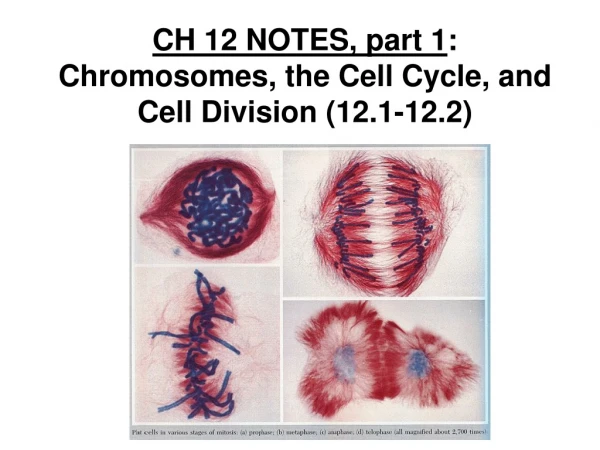
Mitosis is one, continuous event, but it can be described as happening in 5 phases: 1) Prophase 2) Prometaphase 3) Metaphase 4) Anaphase 5) Telophase (followed by CYTOKINESIS!)
G2 OF INTERPHASE PROPHASE PROMETAPHASE
10 µm TELOPHASE AND CYTOKINESIS METAPHASE ANAPHASE
PROPHASE ● chromatin condenses & chromosomes become visible; ● centrosomes / centrioles separate and move to the opposite sides of the nucleus
PROMETAPHASE ●nuclear envelope breaks down and the nucleolus disappears; ● spindle fibers (from centrioles of centrosomes) connect to chromosomes at their centromeres (KINETOCHORE)
METAPHASE ● chromosomes line up in the center of the cell (metaphase plate); ● spindle fibers connect from the poles (end) of the spindle to the centromere / kinetochore of each chromosome
ANAPHASE ● centromeres split, causing the sister chromatids to separate, becoming individual chromosomes ● chromosomes are pulled apart to opposite ends of the cell as the spindle fibers shorten and “reel them in” to the poles
TELOPHASE ● chromosomes uncoil into chromatin; ● new nuclear envelope forms around the chromatin; ● spindle breaks apart; ● nucleolus reappears in each new nucleus
Finally… CYTOKINESIS ● in animal cells: cell membrane pinches in & divides (cleavage furrow) ● in plant cells: a cell plate (new cell wall) forms
100 µm Cleavage furrow Daughter cells Contractile ring of microfilaments Cleavage of an animal cell (SEM)
Vesicles forming cell plate Wall of parent cell 1 µm New cell wall Cell plate Daughter cells Cell plate formation in a plant cell (TEM)
Then the cell returns to Interphase… and the process continues One More Time!
The Mitotic Spindle: A Closer Look ● The mitotic spindle is an apparatus of microtubules that controls chromosome movement during mitosis ● Assembly of spindle microtubules begins in the CENTROSOME, the microtubule organizing center ● The centrosome replicates, forming two centrosomes that migrate to opposite ends of the cell, as spindle microtubules grow out from them ● An aster (a radial array of short microtubules) extends from each centrosome
● The spindle includes: the centrosomes, the spindle microtubules, and the asters ● Some spindle microtubules attach to the kinetochores of chromosomes and move the chromosomes to the metaphase plate
Aster Centrosome Sister chromatids Metaphase plate Chromosomes Microtubules Kineto- chores Overlapping nonkinetochore microtubules Kinetochore microtubules Centrosome 1 µm 0.5 µm
● In anaphase, sister chromatids separate and move along the kinetochore microtubules toward opposite ends of the cell ● The microtubules shorten by depolymerizing at their kinetochore ends
Chromosome movement Kinetochore Tubulin subunits Motor protein Microtubule Chromosome
● Nonkinetochore microtubules from opposite poles overlap and push against each other, elongating the cell ● In telophase, genetically identical daughter nuclei form at opposite ends of the cell
Chromatin condensing Nucleus 10 µm Chromosomes Cell plate Nucleolus Prometaphase. We now see discrete chromosomes; each consists of two identical sister chromatids. Later in prometaphase, the nuclear envelope will fragment. Prophase. The chromatin is condensing. The nucleolus is beginning to disappear. Although not yet visible in the micrograph, the mitotic spindle is starting to form. Metaphase. The spindle is complete, and the chromosomes, attached to microtubules at their kinetochores, are all at the metaphase plate. Telophase. Daughter nuclei are forming. Meanwhile, cytokinesis has started: The cell plate, which will divide the cytoplasm in two, is growing toward the perimeter of the parent cell. Anaphase. The chromatids of each chromosome have separated, and the daughter chromosomes are moving to the ends of the cell as their kinetochore micro- tubules shorten.
BINARY FISSION ● Prokaryotes (bacteria and archaea) reproduce by a type of cell division called BINARY FISSION ● In binary fission, the chromosome replicates (beginning at the origin of replication), and the two daughter chromosomes actively move apart


