Download
1 / 47
1.02k likes | 2.73k Vues
Respiratory System Histology. Dr. Nabil Khouri MD, MSc . Ph.D. Lung Conducting portion INTRAPULMONARY BRONCHI BRONCHIOLES TERMINAL BRONCHIOLES Respiratory portion RESPIRATORY BRONCHIOLES ALVEOLAR DUCTS ALVEOLI. Airways – conducting portion Nasal cavity

E N D
Respiratory System Histology Dr. NabilKhouri MD, MSc. Ph.D
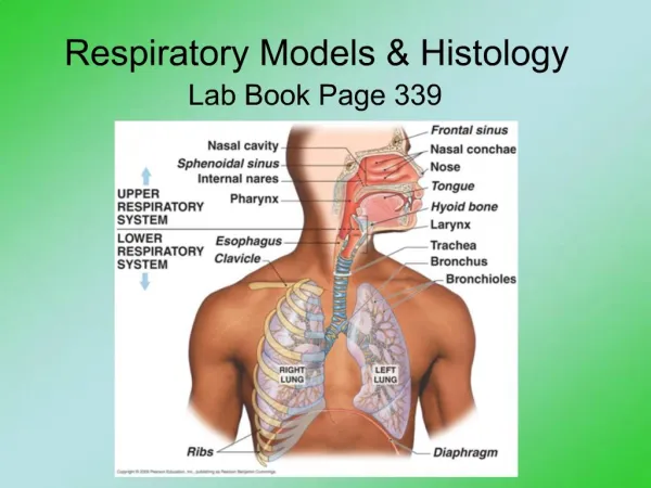
Lung Conducting portion INTRAPULMONARY BRONCHI BRONCHIOLES TERMINAL BRONCHIOLES Respiratory portion RESPIRATORY BRONCHIOLES ALVEOLAR DUCTS ALVEOLI Airways – conducting portion Nasal cavity Pharynx (naso – oro –laringo-) Larynx Trachea Bronchi (extra pulmonary) Respiratory system
Conducting portionNasal Cavity • Nasal cavity composed of three regions( vestibule, respiratory and olfactory regions) • Anterior portion – Vestibule: vibrissae – skin - sebaceous + sweat glands • Posterior aspect – nasal fossae: conchae (sup, mid, inf), olfactory region, • arterial plexuses and venous sinuses bleeding
Vestibule Anterior portion of nasal cavity near of the nares dilated and known as the vestibule • lined with skin and has short, stiff hairs named VIBRISSAE that prevent large dust particles to enter • It is made of Keratinized stratified squamous epithelium And Non Keratinized stratified squamous epithelium • Dermis houses numerous sebaceous glands
Posterior aspect of Nasal cavity • Is lined by Pseudostratified ciliated columnar epithelium (RESPIRATORY EPITHELIUM) • Cellular content of the respiratory epithelium • Ciliated columnar cells: Most common, each cell has about 300 cilia • Goblet cells: Secret mucous (Found in frequency) • Brush cells: Have short microvilli, nerve fibers, sensory function • Basal cells: Are rounded STEM CELLS that located near basal lamina and show mitotic figures • Small granule cells
The Lamina Propria (Subepithelial CT) • Mucous Glands • Serous Glands • Venous Sinuses • lymphoid elements • CT Lamina Propria Is richly vascularized,containing large arterial plexuses and venous sinuses, many • Secretions of nasal mucosa - Bactericides - Lysozymes
Olfactory region • The olfactory epithelium • Lamina propria (serous secreting BOWMAN’S GLANDS (Boumans GL) a rich vascular plexus and many axons arising from olfactory cells of the olfactory epithelium)
olfactory epithelium comprises three types of cells: • Olfactory cells olfactory cells are bipolar neurons whose apical aspect (dendrite) is modified to form a bulb known as olfactory vesicle • Sustentacular cells (Supporting Cells) • Cells that has a striated border composed of microvilli, and secretory granules • They provide physical support, nourishment • Basal cells • Are short basophilic cells • Their apical aspects do not reach the epithelial surface they proliferate and replace both two other cells
Olfactory Epithelium (bipolar neurons) (Sustentacular cells)
The naso-pharynx • The upper part of the pharynx which – • Anteriorly is open into the nasal cavity through posterior nares,choana! • Posteriorly and laterally is surrounded by the muscular tissue • caudally is continued with the oropharynx and larynx • the epithelium of the pharynx is PSEUDOSTRATIFIED CILIATED COLUMNAR • goblet cells and small glands (mucous, serous and mixed) • Accumulations of lymphatic tissue • pharyngeal tonsil – posteriorly • Tubal tonsils – laterally both are found in the lamina propria • The middle layer of the pharynx is of muscular character being formed by bundles of the striated muscles
Pharynx • Oro-pharynx • It is lined by a STRATIFIED SQUAMOUS NON-KERATINIZING TYPE OF EPITHELIUM and lacks both muscularis mucosae and submucosa. • The epithelium rests on a lamina propria that contains a thick layer of longitudinally oriented elastic fibers that appear dark, glassy red located near the underlying muscularis externa. • Lamina propria – loose-dense irregular CT • Vascularized • Seromucous gland: Mucous glands seen in this muscular layer in some of our glass slides are the extensions of those present in the lamina propria. • Lymphoid tissue – posterior: pharyngeal tonsil • Skeletal muscle-epimysium • The muscularis externa is composed of somewhat irregularly arranged skeletal muscle, the longitudinal and constrictor muscles of the pharynx.
Larynx – voice box • Additional function: • Phonation • Prevent food/drinks – respiratory system • Tube : cartilage (hyaline, elastic) – ligaments – skeletal muscles (intrinsic-extrinsic) • Epiglottis – elastic cartilage • Stratified squamous non keratinized epithelium • Pseudostratified (respiratory epithelium) • Vestibular fold – false vocal cord (superior) • Vocal fold - true vocal cord (inferior) - stratified squamous • Vocalis muscle • vocal ligament – regular dense elastic CT
Epiglottis • is an elastic cartilage of larynx • It is lined by stratified squamous epithelium on lingual surface • Pseudostratified ciliated columnar epithelium lined the laryngeal side • Serous and mucous glands located in lamina propria
Trachea–extrapulmonary bronchus • Mucosa • Respiratory epithelium – Pseudostratified ciliated columnar epithelium: goblet, cilated columnar, basal, brush, serous, DNES cells • Lamina propria – loose fibroelastic-mucous, seromucous gl-lymphoid tissue --elastic lamina • Submucosa-dense irreg. fibroelastic CT, mucous-seromucous gl- lymphoid tissue • Adventitia – fibroelastic CT - C ring hyaline cartilage (p cartilaginea) – fibrous CT – smooth muscle (p. membranacea)
Lamina propria composed of loose fibroelastic CT, contain seromucous glands and lyphoid elements, elastic lamina separate this layer from submucosa Submucosa Subnucosa is composed of dense irregular fibroelastic CT that houses mucous and seromucous glands, rich in blood and lymph supply Adventitia Adventitia is a fibroelastic CT that houses C-shaped hyaline cartilage, at posterior aspect of cartilage, there is a dense band of smooth muscle cells known as trachealis muscle
Trachea • The Mucosa • Respiratory epithelium composed of 6 cell types located on a thick basement membrane • Goblet cells are about 30% of cells, produce mucinogen • Ciliated columnar cells about 30% of cells, are tall which have cilia and microvilli • Basal cells are also about 30% of cells, they are undifferentiated stem cells • Brush cells are just 3% of cells, they are narrow columnar cells that their function is unknown, but nerve ending associated with them • Serous cells are about 3% of cells, they are columnar and have serous granule • DNES cells, constitute about 3-4% of cells, have numerous granule in basal cytoplasm which is contain various pharmacological agents
Lung • Intrapulmonary bronchus (2ndary -3tiary: lobe – broncho pulmonary segment) • Mucosa –folded app • Respiratory epithelium • L propria – fibroelastic-seromucous gl-lymphoid nodules --Smooth muscles-spiral • Submucosa-seromucous gl-lymphoid nodules • Adventitia – plates of cartilage (hyaline) • Bronchioles • Mucosa • Pseudostratified columnar epithelium- goblet cells ciliated simple columnar ciliated cuboidal (with Clara cells) • Lamina propria • No glands • Elastic fibers • Smooth muscles – helical loose meshwork – surrounded by fibroelastic CT
Bronchial Tree is composed of: • 2 primary bronchus that enter lungs • 3 lobar ( secondry) bronchus on right and 2 on the left • Segmental (tertiary) bronchus • bronchioles Terminal bronchioles Respiratory bronchioles Progressively airways decreased in size and cartilage, glands, goblet cells, and the height of epithelial cells But increase smooth muscle cells and elastic tissue
Primary Bronchi (Extrapulmonary) PB • Primary bronchi is identical to trachea, but have smaller diameter and thinner wall • Cartilage is in form of irregular plates • Smooth muscle located between lamina propria and submucosa as 2 distinct layers
Bronchioles • Terminal bronchioles are terminus of conducting portion • they are lined by cuboidal cells(some with cilia) and Clara cells which have domed apical surface • Lamina propria • is a fibroelastic CT, 1-2 layer of smooth muscle cells separate it from adventitia • CLARA CELLS ( exocrine bronchiolar cells) • Clara cells are columnar with dome-shaped apex • Secretory granules which secret glycoproteins and surfactant-like materials • degrade toxins(SER) • divide to replace other cells • antimicrobial peptide have no any cartilage or glands but have few Goblet cells In larger bronchioles epithelium is simple columnar ciliated, with occasional goblet cells In smaller bronchioles epithelium change to Simple cuboidal, with no goblet cells Bronchioles have a smooth muscle coats surrounded by fibroelastic connective tissue
Respiratory Bronchioles are a transitional zone between conducting and respiratory tissues Alveoli branching from their walls are lined by ciliated cuboidal epithelium with Clara cells that change to type I alveolar cells Smooth muscle cells and elastic fibers underlie epithelium
Alveolus • Small air sac - gas exchange • Between alveoli – interalveolar septum – alveolar pores • Connective tissue: elastic, reticular (coll III) – lymphoid tissue • Macrophages, fibroblast, myofibroblast, mast cells • Continuous capillary bed (from pulmonary artery vein) • Both side • Type I Pneumocytes/alveolar cells - squamous alveolar cells) – tight junction – basal lamina – very thin region permeable to gasses • Type II Pneumocytes/alveolar cells - great alveolar cell – septal cells – surfactant – surface tension↓ ≠ collapse
Cells of the Alveolar Septa Endothelial cells are nonfenestrated with a thin dark nucleus, and pinocytotic vesicles Type I squamous cells that cover most of alveolar surface area, they have pinocytotic vesicles Type II (greater alveolar) cells are cuboidal, located on alveolar surface where septa intersect, they have foamy cytoplasm, surfactant granules (reduces surface tension to keep alveoli open during expiration) Alveolar macrophages that are known as dust cells Interstitial cells consist of fibroblasts and mast cells Elastic & reticular fibers
Type I pneumocytes (Squamus alveolar cells) • 95% of the alveolar surface is composed of the simple squamous cells which are known as type I pneumocytes • occluding junction attaches to other cells • have basal lamina, • alveolar pore formed by fusion of two adjacent type I cells Type II pneumocytes (Septal cells) • They are more numerous than type I • cover just 5% of the alveolar surface • located among type I cells, cuboidal with dome-shaped apical • Located where adjacent alveoli separated by septum • They have an abundance of RER, developed golgi complex, their lamellar bodies contain pulmonary surfactant
Type II Alveolar or Type II pneumocytes • Also known as Septal cells • Rounded or cuboidal secretory cells with microvilli • Secretory granules are made of several layers- Multilamellar bodies. • These lamillar bodies are cytoplasmic inclusions made up of phospholipid which combines with other chemicals to form surfactant & then ooze out of the cell by exocytosis. • Pulmonary Surfactant – is the fluid secreted that spreads over the alveolar surface • These cells can multiply to replace damaged cells. • Surfactant also has bactericidal properties
Alveolar Macrophages or Dust cells • Derived from Monocytes and are part mononuclear phagocytic system. • Either seen in the septa or alveoli • Cytoplasm contains phagocytosed inhaled carbon and dust particles • Inhaled carbon and dust particles are passed on to them from pneumocyte I through pinocytic vesicles
Alveolar Macrophages or Dust cells • Migrate from septum to alveolar surface and are carried to the pharynx through sputum • Main function is to clean the alveoli of invading microorganisms and inhaled particulate matter by phagocytosis