Download
1 / 32
320 likes | 498 Vues
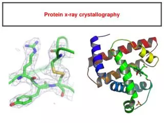
The phase problem in protein crystallography. The phase problem in protein crystallography. Bragg diffraction of X-rays (photon energy about 8 keV, 1.54 Å). Structure factors and electron density are a Fourier pair.

E N D
The phase problem in protein crystallography
The phase problem in protein crystallography
Bragg diffraction of X-rays (photon energy about 8 keV, 1.54 Å)
Structure factors and electron density are a Fourier pair
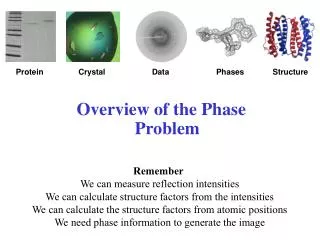
The problem is that the raw data are the squares of the modulus of the Fourier transform. That´s the famous phase problem.
In protein crystallography, there are several ways to get the phases: • Molecular replacement • Heavy atom methods • Direct methods • Non-standard methods
Molecular replacement Mol A: GPGVLIRKPYGARGTWSGGVNDDFFH... Mol B: GPGIGIRRPWGARGSRSGAINDDFGH... ? Mol A Mol B
If we have phases from a similar model... Amplitudes: Manx Phases: Manx Amplitudes: Cat Phases: Cat Phases: Manx Amplitudes: Cat ...we can combine them with the experimental amplitudes to get a better model. we can use
Patterson maps can be used to find .... the proper orientation (rotation) .... the proper position (translation) for the search model. The density map The Patterson map
The Patterson map is the Fourier transform of the intensities. It can be calculated without the phases.
The matching procedure requires a search in up to six dimensions • Luckily, the problem can be factorized into • first, a rotation search • then, a translation search
Flow chart of a typical molecular replacement procedure (AMORE) rotfun (clmn) sortfun hklin (*.mtz) hklpck0 (*0.hkl) clmn0 (*0.clmn) } rotfun (cross) rotfun (generate) rotfun (clmn) tabfun xyzin1 (*1.pdb) table1 (*1.tab) hklpck1 (*1.hkl) clmn1 (*1.clmn) fitfun (rigid) pdbset trafun (CB) rotfun (cross) SOLUTF SOLUTTF solution.pdb SOLUTRC
Poor phases yield self-fulfilling prophesies Amplitudes: Karlé Phases: Karlé Amplitudes: Hauptmann Phases: Hauptmann Amplitudes: Hauptmann Phases: Karlé If Karlé phases Hauptmann, Hauptmann is Karléd!
Can we do holography with crystals? In principle yes, but the limited coherence length requires a local reference scatterer.
For a particular h,k,l FH2 FP FPH1 FH1 FPH1 we can collect all knowledge about amplitudes and phases in a diagram (the so-called Harker diagram)
Normally, there´s the problem that different crystals are not strictly isomorphous. • Thus, the best is a reference scatterer that can be switched on and off.
Absorption is accompanied by dispersion. This Kramers-Kronig equation is very general: Its (almost) only assumption is the existance of a universal maximum speed (c) for signal propagation.
Which elements are useful for MAD data collection? 25 keV 0.5 Å LIII 64- 7 keV 1.8 Å K 26-46
The MAD periodic table H He Li Be B C N O F Ne Na Mg Al Si P S Cl Ar K Ca Sc Ti V Cr Mn Fe Co Ni Cu Zn Ga Ge As Se Br Kr Rb Sr Y Zr Nb Mo Tc Ru Rh Pd Ag Cd In Sn Sb Te I Xe Cs Ba La Hf Ta W Re Os Ir Pt Au Hg Tl Pb Bi Po At Rn Fr Ra Ac Rf Ha Lanthanides Ce Pr Nd Pm Sm Eu Gd Tb Dy Ho Er Tm Yb Lu ActinidesTh Pa U Np Pu Am Cm Bk Cf Es Fm Md No Lr
All phasing can be done on one crystal. F1,2 a b F-1,-2 F1,2 : scattering from b is phase behind F-1,-2 : scattering from b is phase ahead In the presence of absorption, Bijvoet pairs are nonequal.
assuming with absorption:
Direct methods ? Atomic resolution data the best approach for small molecules
If atoms can be treated as point-scatterers, then if all involved structure factors are strong
100 atoms in the unit cell a small protein The method is blunt for lower resolution or too many atoms.
Three-beam phasing ? very low mosaicity data an exciting, but not yet practical way
An example from our work (solved by a combination of MAD and MR) Metal ions
Can we tell from the fluorescence scans? Compton Zn Cu Fe Ni Co Normally yes, but not in this case!
Can we tell from the anomalous signal? order in the periodic table: Fe, Co, Ni, Cu, Zn
Here´s the maps! 2fo-fc map, 1.05 Å anomalous map, 1.05 Å anomalous map, 1.54 Å Quantitatively: f“ (1.05 Å) = 1.85 0.05 f“ (1.54 Å) = 2.4 0.2
Thanks to my group, particularly S. Odintsov and I. Sabała Thanks to Gleb Bourenkov, MPI Hamburg c/o DESY