Understanding the Role of Basal Ganglia in Voluntary Movement Control
The basal ganglia, a cluster of interconnected brain structures, play a crucial role in controlling voluntary movements by regulating motor signals from the brain to the body. They participate in both the initiation and termination of movement by suppressing unwanted actions while facilitating desired ones. This dynamic process involves signals flowing from the motor cortex to the striatum, and then to the globus pallidus and substantia nigra. By understanding this complex network, we can better appreciate how our brain orchestrates movement.


Understanding the Role of Basal Ganglia in Voluntary Movement Control
E N D
Presentation Transcript
Basal Ganglia Animation May 2012
Movement in the body is controlled by the brain. Working in conjunction with motor neurons (neurons that connect to muscles), the brain sends signals to muscles to contract. A combination of these signals results in either a wanted movement or the stopping of an unwanted movement. Highlight first Brain Make shirt solid (not see through) Highlight second Show signal going from brain to hand (brain to motor neurons to hand) Motor Neurons Show movement when “signal” reaches the hand End arrow here
The motor cortex sends out signals to control voluntary movements in the body via motor neurons that cause muscle to contract. Motor Cortex Highlight first Make shirt solid (not see through) Show signal going from brain to hand (motor cortex to motor neurons to hand) Highlight second Motor Neurons Show movement when “signal” reaches the hand
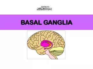
Motor Cortex Highlight first Highlight second The control of voluntary movement by the motor cortex, however, is regulated by other brain circuits. One of these is called the basal ganglia, a group of interconnected structures in the middle of the brain. Specifically, the basal ganglia participate in the initiation and termination of voluntary movements by suppressing unwanted movements and permitting desired and appropriate movements . Highlight third Make shirt solid (not see through) Basal Ganglia Highlight fourth Show signal going from brain to hand (motor cortex to spinal cord, spinal cord to motor neurons, motor neurons to arm and hand muscles AND back and forth between motor cortex and basal ganglia) Motor Neurons Show movement when “signal” reaches the hand
Transition from prior screen: Zoom in on brain and fade basal ganglia area to the actual structures Delete this Basal Ganglia Undo all label lines
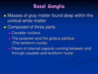
The basal ganglia consists of the striatum (caudate nucleus and putamen), Highlight these Striatum
global pallidus (external and internal segments), Highlight these
subthalamic nucleus, Even though I “cut” out the structures, I would recommend just fading them and highlighting the structure being addressed. Highlight this
and substantianigrareticulata. Highlight these (top half only)
The basal ganglia receives information from most parts of the cerebral cortex and sends information specifically to the motor cortex, to execute movement, through the thalamus. Highlight this
So, how does this process work? Messages are sent from various parts of the cerebral cortex to the basal ganglia, here let’s consider messages from the motor cortex to the striatum, the input structure of the basal ganglia. Motor Cortex Striatum Fill in motor cortex Highlight first Striatum Highlight second Highlight third Keep the highlighting cycling until student moves on to next section.
The basal ganglia also has output structures, the globuspallidus internal segment and the substantianigrareticulata, which control the motor cortex through the thalamus. (N.B. erase big arrow and unhighlight the striatum and highlight the SNr and Gpi as output structures with an arrow going to the thalamus and another arrow going to the motor cortex.) High light first
The globuspallidus internal segment and the substantianigrareticulata control the motor cortex through the thalamus. 4. Highlight this Third – make this a dynamic, dashed line. 2. Highlight second. 1. Have these be dynamic, dashed arrows that appear first and then highlight the thalamus.
The striatum controls the output structures through two different pathways. Striatum Indirect Pathway: Suppresses unwanted or inappropriate movements. Global pallidus external segment Direct Pathway: Promotes wanted or desired movement Subthalamic Nucleus Global pallidus internal segment/Substantianigrareticulata
In the indirect pathway, the striatum sends a message to the global pallidus external segment . Striatum Highlight first Global pallidus external segment Striatum Highlight second Global pallidus external
The global pallidus external then sends a signal to the subthalamic nucleus. Striatum Highlight first Global pallidus external segment Subthalamic nucleus Global pallidus external Subthalamic nucleus Highlight second
The subthalamic nucleus then sends a message to the global pallidus internal and substantianigra. Striatum (Input) Global pallidus external segment Subthalamic nucleus Highlight first Global pallidus internal segment/Substantianigrareticulata (Output) Subthalamic nucleus Global pallidus internal segment Highlight second Substantianigrareticulata
The striatum also sends messages through the direct pathway. In this pathway, the striatum sends a message directly to the global pallidus internal and the substantianigra. Highlight first Striatum Global pallidus internal segment/substantianigrareticulata Hightlight second
Now the global pallidus internal and the substantianigra have received messages from both the indirect and direct pathway. Motor cortex Striatum Global pallidus external Direct Pathway Indirect Pathway Subthalamic nucleus Global pallidus internal/Substantianigra
The global pallidus internal and substantianigra then send a message on to the thalamus. Motor cortex Striatum Highlight first Direct pathway Indirect pathway Global pallidus internal Global pallidus internal/Substantianigra Substantianigra Thalamus Highlight second Thalamus
Motor cortex The thalamus then sends a message t o the motor cortex. Highlight second Striatum Motor Cortex Indirect pathway Direct pathway Global pallidus internal/Substantianigra Thalamus Thalamus Highlight first
The motor cortex then sends the appropriate message out to the body. Highlight first Motor Cortex Highlight second End arrow here