Download
1 / 1
30 likes | 217 Vues
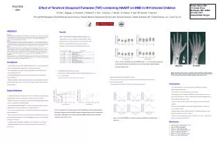
Rohan Hazra, MD 10 Center Drive Bethesda, MD 20892 301-402-1541 hazrar@mail.nih.gov. POSTER 694. Effect of Tenofovir Disoproxil Fumarate (TDF)-containing HAART on BMD in HIV-infected Children

E N D
Rohan Hazra, MD 10 Center Drive Bethesda, MD 20892 301-402-1541 hazrar@mail.nih.gov POSTER 694 Effect of Tenofovir Disoproxil Fumarate (TDF)-containing HAART on BMD in HIV-infected Children RI Gafni1, R Hazra1, JC Reynolds2, F Maldarelli1, A Tullio1, E DeCarlo1, C Worrell1, JF Flaherty3, K Yale3, BP Kearney3, S Zeichner1 1HIV and AIDS Malignancy Branch/National Cancer Institute, 2Nuclear Medicine Department/Clinical Center, National Institutes of Health, Bethesda, MD, 3 Gilead Sciences, Inc., Foster City, CA • ABSTRACT • Background • Tenofovir disoproxil fumarate (TDF), a nucleotide analog HIV reverse transcriptase inhibitor is approved for use in adults but not children. Metabolic bone changes have been seen in young animals given high-dose tenofovir and in HIV-infected adults treated with TDF. Despite concern that children may be at greater risk of bone toxicity than adults, TDF is used off label in pediatrics. We present data on bone toxicity through 96 weeks from a phase 1 study of TDF-containing highly active antiretroviral therapy (HAART) in HIV-infected children. • Methods • Fifteen heavily treatment-experienced HIV-infected children (mean age ± SD, 12 ± 2 y), requiring a change in therapy, received TDF 175-350 mg/m2/d (adult dose equivalent) as part of HAART for up to 96 weeks. Bone mineral density (BMD) of the lumbar spine (LS), femoral neck (FN) and hip (TH) by dual-energy x-ray absorptiometry, metabolic bone markers, and 24 hour urine calcium excretion were measured at 0, 24, 48, 72, and 96 weeks. Comparisons between time points were made using a paired t-test for normally distributed data and a Wilcoxon signed rank test for data not normally distributed. • Results • Baseline mean (± SEM)Z-scores (SD scores compared to age, sex, and ethnicity-matched controls) were below zero (LS -1.02 ± 0.3, TH -0.8 ± 0.4, FN -1.0 ± 0.5).Mean LS, FN, and TH Z-scores decreased further by 24 and 48 weeks and stabilized thereafter in most patients. Absolute decreases in BMD were observed in 6 children, who tended to be younger. The change in LS BMD correlated with decreases in HIV plasma RNA during treatment (r2 = 0.275, P<0.05). Metabolic markers of bone formation and resorption were variable but tended to increase. Twenty-four hour urine calcium excretion was increased at 24 weeks compared with baseline (4.2 ± 1.0 vs. 2.9 ± 0.9 mg/kg/d, P< 0.05). Two children in whom TDF was discontinued at 48 weeks because of significant bone loss demonstrated clear recovery of LS BMD by 96 weeks. • Conclusions • TDF is associated with decreases in BMD in HIV-infected children. Increases in bone markers and calcium excretion suggest that TDF may stimulate bone resorption. Bone turnover is higher in children because of skeletal growth, possibly explaining the greater effect seen in children as compared to adults. While the long-term effects on bone are unknown, careful monitoring of HIV-infected children requiring treatment with TDF is warranted. • Introduction • • Tenofovir disoproxil fumarate (TDF, Viread®, Gilead Sciences, Inc.), a nucleotide analog HIV reverse transcriptase-inhibitor, is approved for use in adults, but not children • • Bone density (BMD) loss is greater in adults receiving TDF-containing HAART than those receiving HAART without TDF1 • • Decreased BMD and bone mineral content have been reported in HIV-infected children treated with HAART2, 3, 4 • • Skeletal abnormalities including defective mineralization, increased resorption cavities, and altered metabolic markers of bone turnover have been reported in young animals treated with high-dose tenofovir 5, 6 • Subjects/Methods • • 15 HAART-experienced HIV-infected children, 8-16 years, received TDF (175-350 mg/m2/d)-containing HAART, as part of an ongoing phase I/II study, for greater than 24 weeks7. • • BMD of the lumbar spine (LS), total hip (TH), and femoral neck (FN) were measured by dual energy x-ray absorptiometry (QDR 4500, Hologic) • • Bone mineral apparent density (BMAD)8, an estimate of volumetric density which is largely independent of bone size, was calculated in the LS: BMAD = BMC/(area)1.5 • • Z-scores (SD score compared to age, sex and ethnicity-matched controls) calculated using published databases 9, 10 • • Markers of bone formation (bone-specific alkaline phosphatase (BSAP) and osteocalcin) and bone resorption (urinary collagen cross-linked N-telopeptide) and urine and serum calcium and phosphorus were measured • In children with open growth plates, radiographs of the left hand and wrist were obtained • • Wilcoxon signed rank test was used to compare values at 24, 48, 72, and 96 weeks to baseline values. Results Table 1.Baseline data for 15 subjects included in analysis. Some subject numbers are missing, reflecting the 4 excluded subjects. Puberty reported as Tanner stage determined by visual inspection of breast and/or pubic hair. Other medications include antiretrovirals and steroid preparations. All steroid therapies were topical, inhaled or intranasal. Figure 2. LS, FN, and TH BMD Z-scores and LS BMAD Z-scores. * P < 0.05 compared to baseline (time 0). Upper and lower borders of the box represent 75th and 25th percentiles respectively. Median indicated by horizontal line. • • Absolute BMD decreases observed in 6 children • • Mean age of these children significantly younger than the BMD stable group [10.2 + 1.1 vs. 13.2 + 1.8 y, p=0.003] • Height and weight Z-scores were stable throughout the study Figure 4. Hand/wrist X-ray of subject 1 at baseline and after 24 weeks of TDF-containing HAART. Cortices appear thinner at 24 weeks and there is decreased new trabecular bone in the areas under the growth plate (primary spongiosa, indicated by arrows). • Markers of bone formation and resorption were variable • 24-hour urine calcium excretion was increased and tubular resorption of phosphorus was decreased at 24 weeks compared to baseline • Two subjects discontinued TDF after 48 weeks due to significant BMD decreases • Conclusions • TDF-containing HAART is associated with decreases in BMD in heavily treatment-experienced HIV-infected children • These decreases were more pronounced in younger children • Discontinuation of TDF in 2 children with BMD loss resulted in improvement at all 3 sites measured (LS, FN, and TH) in 1 child. The other child experienced improved BMD at the LS but further worsening at the other 2 sites. • Increases in calcium excretion and bone markers suggest that TDF may stimulate bone resorption • Decreases in primary spongiosa in some children suggest that bone loss is occurring at areas of rapid bone turnover • Monitoring for bone toxicity in children treated with TDF should be considered (e.g., bone densitometry at baseline and every 6-12 months) • Longer term studies enrolling larger numbers of children are necessary to determine how best to use TDF in HIV-infected children 11 • References • 1. Gilead Sciences, Inc., Viread package insert, 2004. • 2. Mora, et al., AIDS, 15:1823-1829, 2001. • 3. Mora, et al. JCEM, 89: 24-28, 2004. • 4. Arpadi, et al., JAIDS, 29:450-454, 2002. • 5. Tarantal, et al., JAIDS, 29:207-220, 2002. • 6. Castillo, et al., J Orthop Res, 20:1185-1189, 2002. • 7. Hazra, et al., Pediatrics 116:846-854, 2005 • 8. Carter, et al., JBMR, 7:137-145, 1992. • 9. Faulkner, et al., Calcif Tiss Int, 59:344-351, 1996. • Bachrach, et al., JCEM, 84:4702-4712, 1999. • Giacomet, et al., JAIDS, 40:448-450, 2005. Figure 1. Left graph, Percent change in absolute lumbar spine BMD during TDF for individual subjects. Only subjects showing a decrease in BMD at 24 weeks are labeled by subject number, as shown by the filled symbols. Right graph, Percent change in absolute LS BMD from baseline after 24 weeks TDF treatment vs. age. r2=0.215, P = 0.08 Figure 3. LS, FN, and TH BMD in the 2 patients who discontinued TDF after 48 weeks because of bone loss.