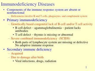
Immunodeficiency
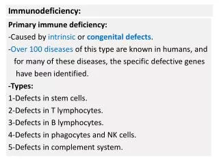
Immunodeficiency. Primary (congenital) immunodeficiencies: inherited disorders that arise from a genetic defect Secondary immunodeficiencies: arise from extrinsic causes Malnutrition Irradiation Infections Immunosuppressive therapy. How Immunodeficiency Diseases Present.

Immunodeficiency
E N D
Presentation Transcript
Immunodeficiency • Primary (congenital) immunodeficiencies: inherited disorders that arise from a genetic defect • Secondary immunodeficiencies: arise from extrinsic causes • Malnutrition • Irradiation • Infections • Immunosuppressive therapy
How Immunodeficiency Diseases Present • Recurrent or chronic pyogenic (pus-forming) infections • These indicate defects in • Antibodies and/or B cells • Complement components • Phagocytes • Common agents are encapsulated bacteria • Streptococcus pneumoniae • Neisseria meningitidis • Staphylococcus aureus • Haemophilus influenzae • Common clinical manifestations • Bacterial pneumonia • Otitis media • Sinusitis • Meningitis • Osteomyelitis • Infections with gram-negative enterics
How Immunodeficiency Diseases Present • Recurrent or chronic opportunistic infections • These indicate defects in T cells • Common agents • Viruses, e.g. rotavirus, HIV • Fungi, e.g. Candida, Pneumocystis jiroveci • Protozoans, e.g. Toxoplasma • Atypical mycobacteria • Leads to combined deficiency of CMI and humoral immunity • Cancer • Usually indicates T cell deficiency • EBV, Kaposi sarcoma
How Immunodeficiency Diseases Present • Diseases that present as antibody deficiencies • X-linked agammaglobulinemia • Selective IgA and IgG subclass deficiencies • Hyper IgM syndrome • Common variable immunodeficiency • Transient hypogammaglobulinemia of infancy • Diseases that present as T cell or combined deficiencies • SCID • DiGeorge syndrome • Hereditary ataxia-telangiectasia • Wiskott-Aldrich syndrome • Phagocytic cell deficiencies • Chronic granulomatous disease • Leukocyte adhesion deficiency • Chédiak-Higashi disease • Hyper IgE (Job) syndrome • Complement deficiencies • Hereditary angioedema • Paroxysmal nocturnal hemoglobinuria • Component deficiencies
X-linked Infantile (Bruton’s) Agammaglobulinemia (XLA) • Males only; rare • btk gene defect • First presents at 5-6 months of age • Repeated pyogenic infections that don’t respond to antibiotics, Giardia lamblia • Passive immunity from mom has dissipated • Near absence of all antibodies: no response to vaccines, no isohemagglutinins • Scant lymphoid tissue • By 20-30 y, bronchiectasis may cause death • T cells OK, so live vaccines OK Antibody deficiencies
Selective IgA Deficiency • Common in Caucasians • Serum IgA <50 mg/ml • Sinopulmonary infections, celiac disease, immune complex disease • May be asymptomatic • May progress to CVID • Persons with IgA deficiency may possess IgE directed toward IgA • Wash those RBCs before transfusion! Antibody deficiencies
Selective IgG Subclass Deficiency • Total serum IgG may be normal • IgG3 deficiency most common in adults • IgG2 deficiency most common in children • May lead to increased bacterial infections, or have no problems Antibody deficiencies
Hyper-IgM Syndrome • Low levels of IgG and IgA with increased levels of IgM • IgM autoantibodies to neutrophils, platelets, tissue antigens • B cells can’t class-switch • X-linked (70%) • T cell defect in CD40L • No T cell activation of macrophages • Defective CMI P. jiroveci • Autosomal recessive (30%) • Defect in CD40 gene in B cells and APCs • Defect in activation-induced cytidine deaminase (AID) gene in the B cell Antibody deficiencies
Common Variable Immunodeficiency (CVID) • Common variable hypogammaglobulinemia • Males and females • Usual onset 15-35 y • May follow EBV infection • Associated with selective IgA deficiency • Low serum IgG and IgA, low or normal IgM, low or normal B cell count • Mothers cannot confer passive protection on infants • Marked by • Pyogenic bacterial infections • Autoimmune diseases, especially pernicious anemia • Giardiasis • Malignant neoplasms • Variable defects Antibody deficiencies
Transient Hypogammaglobulinemia of Infancy • Males and females • Presents at 5-6 months • May persist through 2-3 years of age • IgG absent, IgM and IgA normal • B cells are normal • No class switching to IgG due to TH defect • TH defect precludes live viral vaccines! • Normal numbers of B cells in blood, unlike XLA Antibody deficiencies
Treatments for Antibody Deficiencies • Broad-spectrum antibiotics • Intravenous immune globulin – contains mostly IgG • Do not treat selective IgA deficiency with IVIG • Patients may possess IgE specific for IgA • Administration of IgA may cause anaphylaxis • If your patient is both IgG and IgA deficient, correct the IgG deficiency with an IVIG prep that contains no IgA – Gammagard S/D • Treat IgA deficiency with antibiotics Antibody deficiencies
Severe Combined Immunodeficiency (SCID) • Heterogeneous disorder with defects in CMI and antibody • Patients susceptible to all types of infections – rotavirus, CMV, candidiasis, Pneumocystis jiroveci • Diarrhea and pneumonia • Vaccination with live microbes is lethal • Symptoms occur earlier than in XLA • Fatal before age 1 year if untreated • Treat with bone marrow transplant • There are three phenotypes: T–B+, T–B–, T+B+ T/combined
Severe Combined Immunodeficiency (SCID) • T–B+ subgroup • X-linked • Defect in the g chain of the receptor for IL-2, 4, 7, 9, and 15 • Recessive mutation – heterozygous females are phenotypically normal carriers Failure to activate transcription of specific genes T/combined
Severe Combined Immunodeficiency (SCID) • T–B+ subgroup • Autosomal recessive • Defect in the chain of IL-7 or in the JAK3 tyrosine kinase • JAK3 tyrosine kinase is responsible for transmitting signals from the chain of the receptors for IL-2, 4, 7, 9, 15 T/combined
SCID continued • T-B- subgroup • Defects in adenosine deaminase (ADA) • Accumulation of intracellular S-adenosylhomocysteine and dATP • T and B lymphocytes are particularly susceptible • Lack compensatory 5’-nucleotidase • Treat with gene therapy, continuous enzyme supplementation, or bone marrow transplant • RAG-1 and RAG-2 deficiencies • Stops B and T maturation at pre-B and pre-T stages T/combined
SCID continued • T+B+ subgroup • Bare lymphocyte syndrome • Cells lack MHC class II • No collaboration between APCs and TH cells • Class I MHC/TAP deficiency • Defect in TAP transporter gene • No loading of processed peptide into MHC class I groove • Empty MHC class I molecule is unstable and not expressed on APC surface T/combined
Congenital Thymic Aplasia (DiGeorge Syndrome) • Thymus (and parathyroid) fails to develop from 3rd & 4th pharyngeal pouches • Few or no T cells • Hypocalcemic tetany 24 h due to hypoparathyroidism • Congenital defects in heart and kidneys • Hypertelorism, low-set ears, shortened philtrum T/combined
DiGeorge Syndrome continued • B cells present, but no IgG production • No live vaccines! • Not hereditary, deletion in chromosome 22 • Immunodeficiency treated by bone marrow transplant • BMT may not be necessary • T cell function may be normal by age 5 years • Nude mouse model T/combined
Hereditary Ataxia-Telangiectasia • Neurologic, immunologic, endocrine, hepatic and cutaneous abnormalities • Defective DNA repair gene • DNA breaks in chromosomes 7 and 14 encoding TCR and heavy chain go unrepaired • Presents at 18 months: wobbly gait (ataxia) • Telangiectasia (dilated capillaries) appear on skin and in eyes by 6 y • Severe sinus and lung infections • Autoimmune disorders and cancer T/combined
Wiskott-Aldrich Syndrome • X-linked • wasp gene defect coding for cytoskeletal protein • Decreased signal transduction through adapter proteins • Accelerates Fas-mediated lymphocyte apoptosis • Presents at ~20 months • Thrombocytopenia leading to bleeding disorder • Small platelets • Low IgM, normal IgG, high IgA and IgE • Eczema • Pyogenic and opportunistic infections • Cannot respond to polysaccharide vaccines • Tx: antibiotics, anti-virals, bone marrow transplant • Life expectancy 3 y without antibiotics, 30 y with antibiotics but without immune reconstitution • Death due to lymphoid malignancy T/combined
Chronic Granulomatous Disease (CGD) • Phagocytes lack functional NADPH oxidase no superoxide, no peroxide, leads to intracellular survival of fungi and bacteria • Usual onset early childhood • 2/3 of cases are X-linked • Signs and symptoms • Repeated infections by catalase positive bacteria and fungi • Staphylococcus, Serratia, Burkholderia, Aspergillus, Candida • Granulomas • Pneumonia • Lymphadenitis • Abscesses in skin, liver and viscera • Tx • Antimicrobials, aggressive immunization • IFN • NBT test for diagnosis • CGD phagocytes fail to reduce NBT to formazan • In CGD phagocytes, there’s no color change from yellow to purple Normal CGD Phagocyte defects
Leukocyte Adhesion Deficiency • LAD I • Defective CD18 (an integrin b chain) common to LFA-1 and CR3 • No extravasation • Leukocytosis • No pus formation • No phagocytosis of microbes opsonized by complement • Severe infections spread rapidly, especially in mouth and GI tract • Treat with bone marrow transplant CD15 • LAD II • – Impaired fucose metabolism, e.g. sialyl Lewis X/CD15 Phagocyte defects
Chédiak-Higashi Disease • Defective granules in lysosomes and melanosomes • No intracellular killing • Signs and symptoms • Recurrent Staphylococcus, Streptococcus and Pseudomonas infections of skin, lungs, respiratory tract • Lymphoma-like infiltration of organs by leukocytes with giant cytoplasmic granules • Hypopigmentation because of faulty melanosomes • NK cell activity depressed • Tx: antibiotics, some BMT • Poor prognosis Phagocyte defects
Job’s Syndrome (Hyper IgE Syndrome) • Immunologic features • Recurrent boils and cold staphylococcal abscesses • IgE and chronic eczema • Recurrent cystic lung disease with S. aureus and C. albicans • Mucocutaneous candidiasis • Otitis media • Eosinophilia • Non-immunologic features (not always present) • Bone fractures, other skeletal abnormalities • Coarse facial features, e.g. prominent brow • Joint hyperextensibility • Retained primary dentition • Causative effects variable • Tx: antibiotics and anti-fungals • Tx for eczema: corticosteroids, tacrolimus as last resort Phagocyte defects
Hereditary Angioedema (HAE) • Deficiency in C1 esterase inhibitor allows C1qr2s2 and plasmin to function unregulated • Type I (85%) – low levels of normal C1INH • Type II (15%) – normal or high levels of dysfunctional C1INH • Inherited or spontaneous mutations (acquired AE) • Unchecked activation of classical pathway consumes C4 and C2 • Low levels of C4 are diagnostic • Signs and symptoms • Edema caused by C2 product from plasmin cleavage • Local edema of hands, face, arms, legs, genitals, buttocks • Edema of stomach, intestines, bladder causes abdominal pain, constipation or diarrhea, cramps, vomiting • Laryngeal edema can lead to suffocation • Edema may be preceded by tingling and erythema • Edema caused by stress or trauma, e.g. dental procedure Complement defects
Hereditary Angioedema (HAE) • Prophylaxis before dental or surgical procedures • Fresh frozen plasma to replace C1INH • Danazol to increase C4 • Treatment of on-going attack • Maintain airway • Narcotics for abdominal pain • IV fluids for hemodynamic stability Complement defects
Paroxysmal Nocturnal Hemoglobinuria (PNH) • Stem cell defect in GPI anchors of complement regulators that protect human cells • Decay-accelerating factor (DAF) • Homologous restriction factor (HRF) • CD59 • Dark urine due to RBC lysis • Triad of symptoms • Anemia due to RBC lysis • Pancytopenia due to lysis of RBCs, WBCs, and platelets • Thrombosis in large vessels of liver, abdomen, brain, dermis • Thrombosis usual cause of death in PNH patients • Budd-Chiari syndrome – severe abdominal pain, enlarging liver, ascites • Additional signs and symptoms • Weakness, dyspnea, pallor, splenomegaly, iron deficiency, bleeding disorders, renal failure, severe headaches and eye pain (thrombotic vascular occlusion), chronic infections Complement defects
Paroxysmal Nocturnal Hemoglobinuria (PNH) • Median survival 10 years • Variable age at onset • Dx • Flow cytometry to detect DAF and CD59 on peripheral blood cells • Sugar water test • Reduce salt concentration by adding isotonic sucrose • This causes lysis of PNH RBCs • Ham test • Acidify human serum to pH 6.2 • Add RBCs from PNH patient • RBCs will lyse if GPI-deficient • Tx • For hemolysis: prednisone or eculizumab (Mab to C5) • For anemia: iron supplements, packed RBCs, erythropoietin • For thrombosis: heparin for emergencies, then maintain patient on Coumadin anticoagulant • Stem cell transplant for aplastic anemia or leukemia Complement defects
Complement Component Deficiencies • Deficiency in C5-C8 • Increased incidence of Neisseria infections • Vaccination with MCV4 or MPSV4 helps prevent infections • Deficiency in C3, Factor H, Factor I • Recurrent pyogenic bacterial infections • Vaccinate against Neisseria meningitidis and Streptococcus pneumoniae • Deficiency in C1, C2, C4 • Immune complex disease • SLE • Diagnosis of component deficiencies by CH50 assay • Perform dilution series of patient serum • Add serum dilutions to constant amount of sheep red blood cells coated with IgG • The complement titer is the reciprocal of the highest serum dilution to lyse 50% of the RBCs Complement defects
Secondary Immunodeficiencies • Result from extrinsic causes • Poor nutrition is the leading cause world-wide • Cancer therapy, drugs to prevent graft rejection • Inflammatory bowel disease, skin burns – loss of antibodies • Surgical splenectomy • Infections of leukocytes Secondary immunodeficiencies
Secondary Immunodeficiencies • Acquired immunodeficiency syndrome (AIDS) • Caused by human immunodeficiency virus (HIV) • Transmission • Sexual contact • Transfer of blood products • Contaminated needles • Mother to child during pregnancy, birth, or breast-feeding • HIV infects all cells expressing CD4 • TH cells • Monocytes/macrophages • Dendritic cells • Langerhans cells • Microglial cells Secondary immunodeficiencies
Secondary Immunodeficiencies - AIDS • HIV also requires chemokine receptor as co-receptor • M-tropic viruses use CCR5 • Prefer macrophage host cells • M-tropism early in disease • Persons with mutant CCR5 are HIV-resistant • T-tropic viruses use CXCR4 • Prefer TH host cells • May be evolution from M-tropic to T-tropic as gp120 mutates • Early signs and symptoms • Fevers caused by IL-1 production • Night sweats • Diarrhea • Cachexia due to TNF release Secondary immunodeficiencies
Secondary Immunodeficiencies - AIDS • Later • Profound loss of CD4+ TH cells • <200 TH cells/l • TH:Tc reverses to 1:2 • Loss of humoral and cell-mediated immunity • Recurrent/opportunistic infections • Varicella-zoster – shingles • Herpes simplex – genital and anal lesions • Candida albicans – oral thrush • Cryptosporidium, Campylobacter, Salmonella, microsporidia – diarrhea • Pneumocystis, Mycobacterium, fungi – pneumonia • Toxoplasma – brain abscesses • CMV – esophageal ulceration, chorioretinitis, CNS • JC virus – progressive multifocal leukoencephalopathy • Lymphoma (EBV), Kaposi sarcoma (HHV-8), dementia, paralysis Secondary immunodeficiencies
Kaposi’s sarcoma lesions Pneumocystis jiroveci pneumonia Cryptosporidium CT Cerebral toxoplasmosis Small bowel Secondary immunodeficiencies
Secondary Immunodeficiencies - AIDS • No vaccine, no cure • Tx: highly active antiretroviral therapy (HAART) • Combination therapy from these categories • Nucleoside reverse transcriptase inhibitors • Non-nucleoside reverse transcriptase inhibitors • Protease inhibitors • Inhibitors of viral entry – chemokine receptor antagonists, inhibitors of viral-cell membrane fusion • Integrase inhibitors under development – prevent integration of proviral DNA into host cell genome Secondary immunodeficiencies