Immunodeficiency diseases.
Immunodeficiency diseases. Prof. Mohamed Osman GadElRab. College of Medicine & KKUH. Introduction. Immunodeficiency diseases are : A diverse spectrum of illnesses due to various abnormalities of the immune Prevalence :

Immunodeficiency diseases.
E N D
Presentation Transcript
Immunodeficiency diseases. Prof. Mohamed Osman GadElRab. College of Medicine & KKUH.
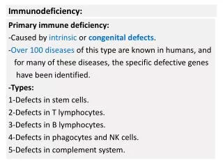
Introduction. • Immunodeficiency diseases are :A diverse spectrum of illnesses due to various abnormalities of the immune • Prevalence : • Primary (congenital) 1 : 10,000 to 1 : 200,000 present at birth . • Secondary (acquired) is more common . MCQ
Overview of Immunodeficiency Disorders. The defect might be In the level of stem cell or in any other level of tree
Clinical manifestations. ( is increased susceptibility to infections ) The patient is considered to have I.D. if the infections are : Frequent & severe. Caused by opportunistic Infections. Resistant to antimicrobial therapy.
Since the main presentation is infection, It is critical to maintain an index of suspicion to diagnose I.D. Important : Early diagnosis & management reduce morbidity (disease).
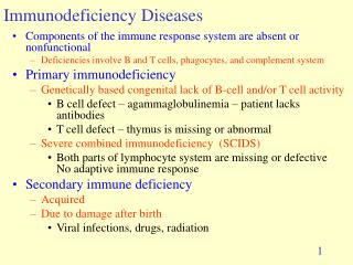
Classification : Primary (congenital). Secondary (acquired). Its common MCQ malnutrition. Most important cause Genetic mutations. Genetic polymorphism. They could be : viral & bacterial infections. e.g. AIDS which is caused by HIV either Monogenic ( defect in one gene ) or polygenic ( defect in more than one gene ) Immunosuppressive drugs. (corticosteroids). For long-time use, it’ll depress the immune system excessive protein loss, burns, nephrotic syndrome ( loss of cells like RBC and loss of protein liek Ig )
Primary or acquired. can affect. Natural immunity (non-specific body defenses). Acquired immunity. (specific body defenses). Phagocytic cells. Complement proteins. T-cells. B-cells.
SCID. Combined T& B cells (SCID). T-cell. T-cell. B-cell. B-cell. Phagocytes. Phagocyte.
The severe combined immunodeficiency is characterized by effecting both B- and T- cells
B-cell defects. Gammaglobulinaemias:
Properties of B-cell defects • Diverse spectrum of diseases ranging from: CompleteabsenceofB-cells , Plasma cells and Immunoglobulin's ,to selective absenceof certain immunoglobulin classes
X- linked disease : • If heterozygous • Female carriers are normal . • Males manifest the disease . • Severity of the disorder parallels ( is Proportional to ) the degree of the deficiency .
] FEATURES OF B-CELL DEFECT [ - Reduced B-cell counts to 0.1 percent ( normally 5-15 percent .) • Absence of Immunoglobulins . • Small Lymph nodes , no germinal centers ( the home of B-cells in the lymph node )
Early B-cell differentiation . Lesions can occur at any site in the pathway of B-cell development. B-cell defect could be in any level in the pathway
IMPORTANTPatients with B-cell defects are subject to: Recurrent bacterial infections but Display normal immunity to most viral & fungal infections. because : T-cells are unaffected. Because T-cell which stands in the face of viral and fungal infection MCQ MCQ
( The disease ) ( the level of defect ) ( the result ) 1. X-linked Bruton tyrosine no mature agammaglobulinaemia. Kinase (Btk) B-cells. Is the first I.D. Recognized in(1952) The most common ( 80 to 90 percent ) Defect in Bruton tyrosine kinase enzyme (BTK). The Defect involve a block in maturation of pre- B- cells to mature B- cells in bone marrow. Pre B-cell + BTK enzyme Mature B-cell MCQ MCQ
Features of XLA.: - Reduced B-cell counts to 0.1 percent ( normally 5-15 percent .) - Absence of Immunoglobulins . - Small L.nodes , no germinal centers . MCQ
X-linked agammaglobulinaemia. (XLA) .cont. . Affected children suffer from recurrent pyogenic bacterial infections of : : (conjunctiva , throat, skin , ear, bronchi & lung) Infecting microbes include :- Pneumococci, H.influenzae Streptococci. Also the patient is susceptible to certain viruses ( polio) and intestinal parasites (giardia ). MCQ
X-linked agammaglobulinaemia. (XLA) .cont. . * Most intracellular microbes & fungi are handled normally by (T- cells ).
2. Selective immunoglobulin deficiency. 1. IgA deficiency (1:700) Most are asymptomatic , but have increased rate of ( respiratory tract infection R.T.I ) Some have recurrent R.T.I. and G.I.T. Symptoms Because of lack of secretion of IgA on the mucous membrane of GIT and respiratory tract . ] Increasedincidence of allergic manifestations [ anti - convlusant drugs (phenytoin) may cause secondary deficiency( these drugs are used to treat epilepsy, they destroy IgA ) MCQ
X- linked hyper-IgM Syndrome. Characterized by : - Low IgG, IgA & IgE - Markedly elevated IgM - High levels of autoantibodies (against neutrophils , platelets , red cells ) ]So, low levels of RBCs, neutrophils, and platelets [ Recurrent infections especially Pneumocystis carinii ]Pneumocystis carinii usually found in people who have AIDS[ MCQ MCQ
X-linked hyper-IgM Syndrome. (cont.) هاااااااام جدا جدا MCQ Defect in the CD 40L in T- cells lead to : ( CD 40L is the hand that Th cell use it to shake other cells hands ) * No co-stimulatory signal for B-cells. * No response to T-dependent antigens . * No class – switching. ( The change of one class of Ig to another one ) * No memory cells. * Marked lymphadenopathy .
( The disease ) ( the level of defect ) ( the result ) 3. X-linked hyper-IgM defective CD40 markedly Syndrome. Ligand. elevated IgM.
Management of immunoglobulin deficiencies : repeated intravenous immunoglobulin (IV Ig) reduces infectious complications . --- GIVE Ig --- MCQ
DiGeorge Syndrome : ( congenital thymic aplasia ) First described in 1952 • Characterized by : - Absence of the Thymus gland . ( So , no T-cells in the body ) - HypoparathyroidismWhich lead to tetany - Cardiovascular abnormalities and Characteristic facial features ]Because they appear from the same embryologic origin of the thymus ( 3-4 pharyngeal pouches) so they are involved[
DiGeorge syndrome : Failure of the third & fourth pharyngeal pouches to develop . *Features : -Children may present with seizures ( tetany) -Extreme susceptibility to viral , protozoal, and fungal infections. ( Because of no T-cell ) So : 1. profound depression of T-cell numbers. 2. absence of T-cell responses. MCQ
DiGeorge syndrome : In some cases B-cells are normal and produce effective humoral immunity to bacterial infections . (Partial Di George Syndrome.) ( thymic hypoplasia, Nezelof syndrome ). There’s a little number of circulating T-cell but B-cells are normal In some T-cell – dependant antibody production is absent .( no helper T- cells ).
DiGeorge syndrome ; • Management: Fetal thymus tissue graft (14 week old). steps should be taken to prevent G.V.H. ( graft versus host ) Reactions G.V.H. Reactions : the transplanted thymus or bone marrow recognize the host body cells as foreign body’s cells and attack it MCQ
Severe combined immunodeficiency. (SCID ). Both T and B cells are defected
Severe combined I.D. : Features: 1. Increased susceptibility to viral, fungal , bacterial & protozoal infection. ( start at 3 month of age ) 2. Failure to thrive. 3. Reduced weight gain. 4. Prolonged diarrhea. 5. Moniliasis due to candida .
Severe combined immunodeficiency (SCID ) : inAutosomal recessive SCID - ADA deficiency . toxic metabolites in T & B-cells. - PNP deficiency. ( ADA and PNP are missing enzymes) MCQ Lead to
Management of recessive (SCID.) 1. Infusion of purified enzymes. 2. Gene therapy . MCQ
Leukocyte defects. Its either : Quantitative. ( amount ) Qualitative. ( function )
Quantitative. 1. Congenital agranulocytosis : ] Kostmann syndrome [ Defect in the gene inducing G-CSF(granulocyte colony stimulating factor)it is a stimulatory factor that acts on bone marrow to produce WBCs, so if its defected, the amount will decrease Features:pneumonia ,otitis media, gingivostomatitis perineal abscesses Management: Respond to G-CSF therapy ( gene therapy ) MCQ
Qualitative. 1.Defect in response to chemotactic agents. 2.Defect in intracellular killing. A . Defect in chemotaxis: Leukocyte adhesion deficiency(LAD.)
B. Defect in intracellular killing: 1.Chronic granulomatous disease: x-linked. (75%) autosomal recessive .(25%). DEFECT: in the oxidative complex . ( responsible for producing superoxide radicals .) FEATURES: Extreme susceptibility to infections. Granulomatous inflammation. (chronic T-cell stimulation.) MCQ
Deficiency of all complement components have been described C1-C9. 1. Deficiency of C1, C2 & C4. ( classical pathway ) lead to immune-complex diseases which can cause significant pathology in autoimmune diseases. ] N.B : immune-complex = antibody - antigen interaction [
antibody independent antibody dependent Activation of C3 and generation of C5 convertase activation Of C5 LYTIC ATTACK PATHWAY Pathways of complement activation. LECTIN PATHWAY ALTERNATIVE PATHWAY CLASSICAL PATHWAY MCQ
The main component that needs to be activated is C3 , All pathways activate C3 • C3 then activates C5 and divide intoC3a and C3b • C5 then activate (membrane - attackcomplex) anddivide intoC5a and C5b • In classic pathway : • C1 activates C2 which activates C4 finally activates C3 MCQ
4. Deficiency of membrane - attack complex. (MAC).( C5 - C9 ) Lead to infection with N.meningitides and N.gonorrhea .
Deficiency of C3 ( the central point of complement ) ] no complement proteins [ • Lead to infections with pyogenic bacteria. • impaired clearance of immune-complexes. . MCQ
C1 - inhibitor deficiency:( hereditary angioedema ) • Is a deficiency of an enzyme which is responsible for prevention of ]C1 self-activation[ • because it’ll attack the body’s own cells, and cause inflammation usually in the ]uvula[ which leads to choking till death. • So these patients always have adrenaline in their pockets to prevent the swallowing • So the enzyme makes C1 activated onlyagainst pathogens.
4. Laboratory evaluation. 1. Complete blood count (total & differential). 2. Evaluation of antibody responses :- A. determination of serum immunoglobulins B. measure specific antibody responses : -To polysaccharide antigens. ( measure isohemagglutinins. ) - To protein antigens . ( measure antibodies to tetanus .)
3. Determination of T & B cell counts.( by flow cytometry ) 4. Determination of the complement components. C3, C4 . - assess functional activity by CH50 5. Assess phagocyte function. - phagocytosis & respiratory burst 6. Carrier detection & prenatal diagnosis ( important for genetic counseling ) MCQ
to know which pathway activates the complement : • we check C2 and C3 levels, if both are decreased, it means they have been used a lot , so it’s the classic pathway • If C3 levels are the only reduced means lactin or alternative pathway.