Download
1 / 29
290 likes | 445 Vues
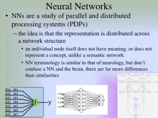
EE 671 – Neural Networks Lecture 1. Nervous System Source: http://www2.estrellamountain.edu/faculty/farabee/biobk/biobooktoc.html. Contents. Neuron Nervous System Brain Spinal Cord Senses Mind Intelligence Consciousness. Nerve Cells.

E N D
EE 671 – Neural NetworksLecture 1 Nervous System Source: http://www2.estrellamountain.edu/faculty/farabee/biobk/biobooktoc.html
Contents • Neuron • Nervous System • Brain • Spinal Cord • Senses • Mind • Intelligence • Consciousness
Nerve Cells • Nerve Cells are of two types: Glial Cell and Neurons • Glial cells protect neurons and do not participate in receiving/transmitting of message • Each Yellow Structure represents a neuron • The neuron is the functional unit of the nervous system • 100 billion (approx) neurons in a human brain
A Neuron • Each neuron has three parts: Dendrites, Cell Body and Axon • Dendrites receive information from another cell and transmit the message to the cell body • The cell body contains the nucleus, mitochondria and other organelles typical of eukaryotic cells. • The axon conducts messages away from the cell body.
Three Types of Neurons • Sensory neurons typically have a long dendrite and short axon, and carry messages from sensory receptors to the central nervous system. • Motor neurons have a long axon and short dendrites and transmit messages from the central nervous system to the muscles (or to glands). • Interneurons are found only in the central nervous system where they connect neuron to neuron.
Nerve Message • resting potential: The difference in electrical charge across the plasma membrane of a neuron. • action potential: A reversal of the electrical potential in the plasma membrane of a neuron that occurs when a nerve cell is stimulated; caused by rapid changes in membrane permeability to sodium and potassium.
Action Potential • The action potential begins at one spot on the membrane, but spreads to adjacent areas of the membrane, propagating the message along the length of the cell membrane. After passage of the action potential, there is a brief period, the refractory period, during which the membrane cannot be stimulated. This prevents the message from being transmitted backward along the membrane.
Steps in an action potential • At rest the outside of the membrane is more positive than the inside. • Sodium moves inside the cell causing an action potential, the influx of positive sodium ions makes the inside of the membrane more positive than the outside. • Potassium ions flow out of the cell, restoring the resting potential net charges. • Sodium ions are pumped out of the cell and potassium ions are pumped into the cell, restoring the original distribution of ions.
Synapse • The junction between a nerve cell and another cell is called a synapse. • Messages travel within the neuron as an electrical action potential. • The space between two cells is known as the synaptic cleft. • To cross the synaptic cleft requires the actions of neurotransmitters. • Neurotransmitters are stored in small synaptic vessicles clustered at the tip of the axon.
Nervous System Three basic functions are performed by nervous systems: • Receive sensory input from internal and external environments • Integrate the input • Respond to stimuli • Sensory Input: Receptors are parts of the nervous system that sense changes in the internal or external environments. Sensory input can be in many forms, including pressure, taste, sound, light, blood pH, or hormone levels, that are converted to a signal and sent to the brain or spinal cord. • Integration and Output: In the sensory centers of the brain or in the spinal cord, the barrage of input is integrated and a response is generated. The response, a motor output, is a signal transmitted to organs than can convert the signal into some form of action, such as movement, changes in heart rate, release of hormones, etc.
Nervous System • Peripheral Nervous System • Somatic Nervous System • Autonomic Nervous System • Central Nervous System
Peripheral Nervous System • The Peripheral Nervous System (PNS)contains only nerves and connects the brain and spinal cord (CNS) to the rest of the body. The axons and dendrites are surrounded by a white myelin sheath. Cell bodies are in the central nervous system (CNS) or ganglia. Ganglia are collections of nerve cell bodies. Cranial nerves in the PNS take impulses to and from the brain (CNS). Spinal nerves take impulses to and away from the spinal cord. There are two major subdivisions of the PNS motor pathways: the somatic and the autonomic. Two main components of the PNS: • sensory (afferent) pathways that provide input from the body into the CNS. • motor (efferent) pathways that carry signals to muscles and glands (effectors). Most sensory input carried in the PNS remains below the level of conscious awareness. Input that does reach the conscious level contributes to perception of our external environment.
Somatic Nervous System • The Somatic Nervous System (SNS) includes all nerves controlling the muscular system and external sensory receptors. External sense organs (including skin) are receptors. Muscle fibers and gland cells are effectors. The reflex arc is an automatic, involuntary reaction to a stimulus. When the doctor taps your knee with the rubber hammer, she/he is testing your reflex (or knee-jerk). The reaction to the stimulus is involuntary, with the CNS being informed but not consciously controlling the response. Examples of reflex arcs include balance, the blinking reflex, and the stretch reflex. • Sensory input from the PNS is processed by the CNS and responses are sent by the PNS from the CNS to the organs of the body. • Motor neurons of the somatic system are distinct from those of the autonomic system. Inhibitory signals, cannot be sent through the motor neurons of the somatic system.
Autonomous Nervous System • The Autonomic Nervous System is that part of PNS consisting of motor neurons that control internal organs. It has two subsystems. The autonomic system controls muscles in the heart, the smooth muscle in internal organs such as the intestine, bladder, and uterus. The Sympathetic Nervous System is involved in the fight or flight response. The Parasympathetic Nervous System is involved in relaxation. Each of these subsystems operates in the reverse of the other (antagonism). Both systems innervate the same organs and act in opposition to maintain homeostasis. For example: when you are scared the sympathetic system causes your heart to beat faster; the parasympathetic system reverses this effect. • Motor neurons in this system do not reach their targets directly (as do those in the somatic system) but rather connect to a secondary motor neuron which in turn innervates the target organ.
Central Nervous System • The Central Nervous System (CNS) is composed of the brain and spinal cord. The CNS is surrounded by bone-skull and vertebrae. Fluid and tissue also insulate the brain and spinal cord.
The Brain • The brain is composed of three parts: the cerebrum (seat of consciousness), the cerebellum, and the medulla oblongata (these latter two are "part of the unconscious brain"). • The medulla oblongata is closest to the spinal cord, and is involved with the regulation of heartbeat, breathing, vasoconstriction (blood pressure), and reflex centers for vomiting, coughing, sneezing, swallowing, and hiccuping. The hypothalamus regulates homeostasis. It has regulatory areas for thirst, hunger, body temperature, water balance, and blood pressure, and links the Nervous System to the Endocrine System. The midbrain and pons are also part of the unconscious brain. The thalamus serves as a central relay point for incoming nervous messages.
Brain • The cerebellum is the second largest part of the brain, after the cerebrum. It functions for muscle coordination and maintains normal muscle tone and posture. The cerebellum coordinates balance. • The conscious brain includes the cerebral hemispheres, which are are separated by the corpus callosum. In reptiles, birds, and mammals, the cerebrum coordinates sensory data and motor functions. The cerebrum governs intelligence and reasoning, learning and memory. While the cause of memory is not yet definitely known, studies on slugs indicate learning is accompanied by a synapse decrease. Within the cell, learning involves change in gene regulation and increased ability to secrete transmitters.
Brain • The Brain Stem: The brain stem is the smallest and from an evolutionary viewpoint, the oldest and most primitive part of the brain. The brain stem is continuous with the spinal cord, and is composed of the parts of the hindbrain and midbrain. The medulla oblongata and pons control heart rate, constriction of blood vessels, digestion and respiration. • Midbrain: The midbrain consists of connections between the hindbrain and forebrain. Mammals use this part of the brain only for eye reflexes. • The Cerebellum: The cerebellum is the third part of the hindbrain, but it is not considered part of the brain stem. Functions of the cerebellum include fine motor coordination and body movement, posture, and balance. This region of the brain is enlarged in birds and controls muscle action needed for flight. • The Forebrain: The forebrain consists of the diencephalon and cerebrum. The thalamus and hypothalamus are the parts of the diencephalon. The thalamus acts as a switching center for nerve messages. The hypothalamus is a major homeostatic center having both nervous and endocrine functions.
Functional areas of the brain • The occipital lobe (back of the head) receives and processes visual information. The temporal lobe receives auditory signals, processing language and the meaning of words. The parietal lobe is associated with the sensory cortex and processes information about touch, taste, pressure, pain, and heat and cold. • The frontal lobe conducts three functions: • motor activity and integration of muscle activity • speech • thought processes
Motor Cortex • Most people who have been studied have their language and speech areas on the left hemisphere of their brain. Language comprehension is found in Wernicke's area. Speaking ability is in Broca's area. Damage to Broca's area causes speech impairment but not impairment of language comprehension. Lesions in Wernicke's area impairs ability to comprehend written and spoken words but not speech. The remaining parts of the cortex are associated with higher thought processes, planning, memory, personality and other human activities.
The Spinal Cord • The spinal cord runs along the dorsal side of the body and links the brain to the rest of the body. Vertebrates have their spinal cords encased in a series of (usually) bony vertebrae that comprise the vertebral column. • The gray matter of the spinal cord consists mostly of cell bodies and dendrites. The surrounding white matter is made up of bundles of interneuronal axons (tracts). Some tracts are ascending (carrying messages to the brain), others are descending (carrying messages from the brain). The spinal cord is also involved in reflexes that do not immediately involve the brain.
Brains and Drugs • Some neurotransmitters are excitory, such as acetylcholine, norepinephrine, serotonin, and dopamine. Some are associated with relaxation, such as dopamine and serotonin. Dopamine release seems related to sensations of pleasure. Endorphins are natural opioids that produce elation and reduction of pain, as do artificial chemicals such as opium and heroin. Neurological diseases, for example Parkinson's disease and Huntington's disease, are due to imbalances of neurotransmitters. Parkinson's is due to a dopamine deficiency. Huntington's disease is thought to be cause by malfunctioning of an inhibitory neurotransmitter. Alzheimer's disease is associated with protein plaques in the brain. • Drugs are stimulants or depressants that block or enhance certain neurotransmitters. Dopamine is thought involved with all forms of pleasure. Cocaine interferes with uptake of dopamine from the synaptic cleft. Alcohol causes a euphoric "high" followed by a depression. • Marijuana, material from the Indian hemp plant (Cannabis sativa), has a potent chemical THC (tetrahydracannibinol) that in low, concentrations causes a euphoric high (if inhaled, the most common form of action is smoke inhalation). High dosages may cause severe effects such as hallucinations, anxiety, depression, and psychotic symptoms. • Cocaine is derives from the plant Erthoxylon coca. Inhaled, smoked or injected. Cocaine users report a "rush" of euphoria following use. Following the rush is a short (5-30 minute) period of arousal followed by a depression. Repeated cycle of use terminate in a "crash" when the cocaine is gone. Prolonged used causes production of less dopamine, causing the user to need more of the drug. • Heroin is a derivative of morphine, which in turn is obtained from opium, the milky secretions obtained from the opium poppy, Papaversomniferum. Heroin is usually injected intravenously, although snorting and smoking serve as alternative delivery methods. Heroin binds to ophioid receptors in the brain, where the natural chemical endorphins are involved in the cessation pain. Heroin is physically addictive, and prolonged use causes less endorphin production. Once this happens, the euphoria is no longer felt, only dependence and delay of withdrawal symptoms.
Senses • Input to the nervous system is in the form of our five senses: pain, vision, taste, smell, and hearing. Vision, taste, smell, and hearing input are the special senses. Pain, temperature, and pressure are known as somatic senses. Sensory input begins with sensors that react to stimuli in the form of energy that is transmitted into an action potential and sent to the CNS. • Sensory Receptors • Sensory receptors are classified according to the type of energy they can detect and respond to. • Mechanoreceptors: hearing and balance, stretching. • Photoreceptors: light. • Chemoreceptors: smell and taste mainly, as well as internal sensors in the digestive and circulatory systems. • Thermoreceptors: changes in temperature. • Electroreceptors: detect electrical currents in the surrounding environment. • Mechanoreceptors vary greatly in the specific type of stimulus and duration of stimulus/action potentials. The most adaptable vertebrate mechanoreceptor is the hair cell. Hair cells are present in the lateral line of fish. In humans and mammals hair cells are involved with detection of sound and gravity and providing balance.
Senses • Hearing: Hearing involves the actions of the external ear, eardrum, ossicles, and cochlea. In hearing, sound waves in air are converted into vibrations of a liquid then into movement of hair cells in the cochlea. Finally they are converted into action potentials in a sensory dendrite connected to the auditory nerve. Very loud sounds can cause violent vibrations in the membrane under hair cells, causing a shearing or permanent distortion to the cells, resulting in permanent hearing loss. • Orientation and Gravity: Orientation and gravity are detected at the semicircular canals. Hair cells along three planes respond to shifts of liquid within the cochlea, providing a three-dimensional sense of equilibrium. Calcium carbonate crystals can shift in response to gravity, providing sensory information about gravity and acceleration.
Photoreceptors Detect Vision and Light Sensitivity The human eye can detect light in the 400-700 nanometer (nm) range, a small portion of the electromagnetic spectrum, the visible light spectrum. Light with wavelengths shorter than 400 nm is termed ultraviolet (UV) light. Light with wavelengths longer than 700 nm is termed infrared (IR) light.
Eye • In the eye, two types of photoreceptor cells are clustered on the retina, or back portion of the eye. These receptors, rods and cones, apparently evolved from hair cells. Rods detect differences in light intensity; cones detect color. Rods are more common in a circular zone near the edge of the eye. Cones occur in the center (or fovea centralis) of the retina. • Light reaching a photoreceptor causes the breakdown of the chemical rhodopsin, which in turn causes a membrane potential that is transmitted to an action potential. The action potential transfers to synapsed neurons that connect to the optic nerve. The optic nerve connects to the occipital lobe of the brain. • Humans have three types of cones, each sensitive to a different color of light: red, blue and green. Opsins are chemicals that bind to cone cells and make those cells sensitive to light of a particular wavelength (or color). Humans have three different form of opsins coded for by three genes on the X chromosome. Defects in one or more of these opsin genes can cause color blindness, usually in males.
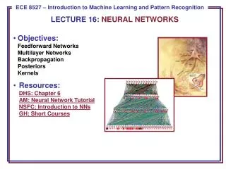
Information Processing in the Brain Neurons typically operate at a maximum rate of about 100 Hz, while a conventional CPU carries out several hundred million machine level operations per second. Despite of being built with very slow hardware, the brain has quite remarkable capabilities: • its performance tends to degrade gracefully under partial damage. In contrast, most programs and engineered systems are brittle: if you remove some arbitrary parts, very likely the whole will cease to function. • it can learn (reorganize itself) from experience. • this means that partial recovery from damage is possible if healthy units can learn to take over the functions previously carried out by the damaged areas. • it performs massively parallel computations extremely efficiently. For example, complex visual perception occurs within less than 100 ms, that is, 10 processing steps! • it supports our intelligence and self-awareness. (Nobody knows yet how this occurs.)
Sample Questions • What tasks are machines good at doing that humans are not? • What tasks are humans good at doing that machines are not? • What tasks are both good at? • What does it mean to learn? • How is learning related to intelligence? • What does it mean to be intelligent? Do you believe a machine will ever be built that exhibits intelligence? • Have the above definitions changed over time? • If a computer were intelligent, how would you know? • What does it mean to be conscious? • Can one be intelligent and not conscious or vice versa?