Download
1 / 54
540 likes | 571 Vues
Learn about the muscles and movements of the knee joint, including ligaments, menisci, and bursae. Understand the function of crucial structures and how they contribute to knee mobility and stability.

E N D
MUSCLES AND MOVEMENTS OF KNEE JOINT MUSCLES AND JOINTS OF THE FOOT ARCHITEXTURE OF THE FOOT Dr. Katalin Gallatz
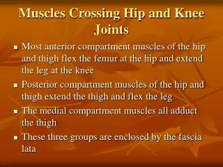
Knee joint: Type:hinge joint + pivot=trochoginglymus Movements: • Flexion, extension- Medial and lateral rotation (only in flexed position!) - bonycomponents - articularcapsule - ligaments - menisci - movements - muscles
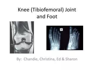
KNEE JOINT Tibiofemoral and patellofemoraljoints Mechanism: trochoginglymus!!! Originally two jointsbetweenthefemur and tibia: - 4 collateral ligaments: medial and lateralcollaterallig. anterior and posteriorcruciatelig. Incongruentsurfaces: equalizedbymed. and lat. meniscus Great workload, frequently damaged. (skiing, football) Movements: flexion:130° ; extension:0-5° ; rotation:50° (only at flexed knee!!!)
LIGAMENTS: - medial (tibial) and lateral (fibular) collaterallig. - anterior and posteriorcruciatelig.
COLLATERAL LIGAMENTS Theystabilizethekneejoint: - theyare strechedonly at extended knee. - inflexed positiontheyarerelaxed and do not limit any movements (rotation is possible!)
ANTERIOR AND POSTERIOR CRUCIATE LIGAMENTS Some parts are streched at every position, Limit medial and lateral rotation, antero-posterior movements.
OTHER LIGAMENTS Patellarligament Obliquepopliteallig. Meniscofemorallig. Transverselig. of knee
PATELLA • It is embeddedintothepatellar ligament, itfunctions • primarilyas an anatomicpulleyforthequadricepsmuscle. • Duringextensionitliesonthepatellarysurface of thefemur, • Duringflexionitmoves down intotheintercondylar fossa whereitliesontheinfrapatellaryfat pad
MENISCI • -The meniscus serves as a shock-absorption system, • - limits the ability to flex and extend the joint. • - equalizetheincongruentsurfaces, • obtain differentpositions at different stages of • movements. • Medial meniscus is more fixed – damaged more often. • Symptoms: pain, „stop of movement” Lateralmeniscus Medialmeniscus Sobota - Atlas of Human Anatomy Passive movements of menisci
INFRAPATELLAR FAT PAD Duringtheflexionthepatellamoves down fromthepatellarysurface of thefemurintotheintercondylar fossa and theinfrapatellarfat pad movesup and fillsthe fossa, providing an adequatesurfaceforthepatella
Theknee bursae are the fluid-filled sacs that surround and sometimes communicate with the knee joint cavity, • present between the skin and tendon or tendon and bone. • They represent the weak point of the joint, but also provide enlargements • to the joint space. • The main function of a bursa is to reduce friction between adjacent • moving structures.
MOVEMENTS Flexion - extension 400 100 „activerotation”
Obligatoryterminal/finalrotation (50) /”passiverotation”/ During the last 10° of extension, an obligatory terminal rotation is triggered in which the knee is rotated medially 5°. • Reasons: • The anteriorcruciate ligament • shorterthantheposterior. • 2) The medialcondylelongerwith • 2 cm thanlateral.
Extended position Inextendedkneeboth the lateral and medial collateral ligaments, as well as the anterior part of the anterior cruciate ligament, istaut. During the last 10° of extension, an obligatory terminal rotation is triggered in which the knee is rotated medially 5°. Flexed position In the flexed position, the collateral ligaments are relaxed while the cruciate ligaments are taut. Rotation is controlled by the twisted cruciate ligaments; the two ligaments get twisted around each other during medial rotation of the tibia, while they become unwound during lateral rotation of the tibia. Because of the oblique position of the cruciate ligaments at least a part of one of them is always tense and these ligaments control the joint as the collateral ligaments are relaxed. Furthermore, the dorsal fibers of the tibial collateral ligament become tensed during extreme medial rotation and the ligament also reduces the lateral rotation to 45–60°.
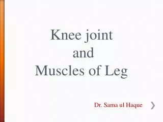
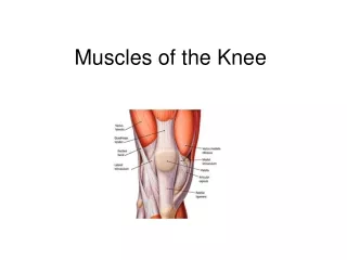
Muscles of thekneejoint: Flexion Semitendinosus m. Semimebranosusm. Bicepsfemoris m. Gracilis m. Sartoius m. Popliteus m. Gastrocnemius m. l. Extension: m. quadricepsfemoris tensorfasciaelates „obligatoryterminalrotation„ Medialrotation: Semimemranosus m. Semitendinosus m. Gracilis m. Popliteus m. Lateralrotation: Bicepsfemoris m. Tensorfasciaelatae
Superficial pes anserinus Common insertionof thesartorius, gracilis, semitendinosusmusclesjust below the medial condyle of the tibia Deep pes anserinus Insertion of thesemimembranosusmuscle, belowthesuperficialpesanserinus „pesanserinus” = „leg of goose”
Popliteal fossa Borders: semitendinosus et semimembranosus m. – SUPERIOR MEDIAL biceps femoris m. – SUPERIOR LATERAL m. gastrocnemius medialis et lateralis –INFERIOR MEDIAL AND LATERAL Base: femur (poplitealsurface), m. adductor magnus, capsule of knee joint, popliteus m. Contents: popliteal artery and vein tibial nerve, common peroneal nerve(sciatic n.), lesser saphenous vein, lymph vessels, med. and lat. cutaneous. sural nerve. (sural n.), collateral arteries/veins of the knee. Sobota - Atlas of Human Anatomy
Thetalocalcaneonavicularjoint is a ball and socket joint: - the rounded head of the talusarticulateswiththe posterior surface of the navicularbone, - the anterior and middlearticular surface of the talus, is connectedwiththeanterior and middlearticularsurface of calcaneus, thesocket made bynavicularbone and calcaneus is completed bytheplantar calcaneonavicular ligament.
MOVEMENTS IN TALOCALCANEONAVICULAR JOINT MOVEMENTS OF THE FOOT MOVEMENTS IN ANKLE JOINT
AXIS OF THE MOVEMENTS (INVERSION – EVERSION) IN TALOCALCANEONAVICULAR JONT AXIS OF THE MOVEMENTS (DORSALFLEXION - PLANTARFLEXION ) IN TALOCRULAR JONT
Hingejoints Restricted ball and socketjoints LISFRANC LINE
FLATFOOT FOOT PRINT
CLINICALLY IMPORTANT LISFRANC LINE TRANSVERSE TARSAL JOINT CHOPART JOINT
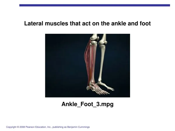
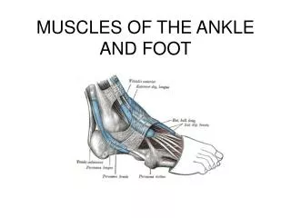
MUSCLES FUNCTION Tricepssuraeplantarflexion, Tibialisposteriorplantarflexion, supination and adduction fibularislongus and brevisplantarflexionandpronation Tibialisanteriordorsalflexion and supination Extensorhallucislongusdorsalflexion and extension of thebigtoe Extensordigitorumlongusdorsalflexion and extension of the 2-4 fingers