Neural Control
Explore the evolution of nervous systems and the impact of drugs like Ecstasy on neural controls. Learn about different types of neurons, nerve nets in animals, and the functioning of sensory, interneurons, and motor neurons. Discover the organization of nervous tissue in animals and how neurons communicate through dendrites and axons.

Neural Control
E N D
Presentation Transcript
Neural Control Chapter 33 Part 1
Impacts, IssuesIn Pursuit of Ecstasy • Neural controls maintain life; drugs like Ecstasy flood the brain with signaling molecules and saturate receptors, disrupting these controls
33.1 Evolution of Nervous Systems • Interacting neurons allow animals to respond to stimuli in the environment and inside their body • Neuron • A cell that can relay electrical signals along its plasma membrane and can communicate with other cells by specific chemical messages • Neuroglia • Support neurons functionally and structurally
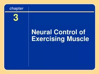
Three Types of Neurons • Sensory neurons detect stimuli and signal interneurons or motor neurons • Interneurons process information from sensory neurons and send signals to motor neurons • Motor neurons control muscles and glands
The Cnidarian Nerve Net • Cnidarians are the simplest animals that have neurons, which are arranged as a nerve net • Nerve net • A mesh of interconnecting neurons with no centralized controlling organ
Bilateral, Cephalized Nervous System • Flatworms are the simplest animals with a bilateral, cephalized nervous system • Cephalization • The concentration of neurons that detect and process information at the body’s head end • Ganglion • A cluster of neuron cell bodies that functions as an integrating center
Nerve Cords • Annelids and arthropods have paired ventral nerve cords that connect to a simple brain • Pair of ganglia in each segment for local control • Chordates have a single, dorsal nerve cord; vertebrates have a brain at the anterior region of the nerve cord
a nerve net (highlighted in purple) controls the contractile cells in the epithelium Hydra, a cnidarian Fig. 33-2a, p. 554
pair of ganglia pair of nerve cords cross-connected by lateral nerves Planarian, a flatworm Fig. 33-2b, p. 554
rudimentary brain ventral nerve cord ganglion c Earthworm, an annelid Fig. 33-2c, p. 554
brain brain optic lobe (one pair, for visual stimuli) branching nerves paired ventral nerve cords ganglion Crayfish, a crustacean (a type of arthropod) Grasshopper, an insect (a type of arthropod) Fig. 33-2 (d-e), p. 554
The Vertebrate Nervous System • Central nervous system (CNS) • Brain and spinal cord (mostly interneurons) • Peripheral nervous system (PNS) • Nerves from the CNS to the rest of the body (efferent) and from the body to CNS (afferent) • Autonomic nerves and somatic nerves control different organs of the body
Functional Divisions of the Vertebrate Nervous System Central Nervous System Brain Spinal Cord Peripheral Nervous System (cranial and spinal nerves) Autonomic Nerves Somatic Nerves Nerves that carry signals to and from smooth muscle, cardiac muscle, and glands Nerves that carry signals to and from skeletal muscle, tendons, and the skin Sympathetic Division Parasympathetic Division Two sets of nerves that often signal the same effectors and have opposing effects Stepped Art Fig. 33-3, p. 555
Major Nerves of the Human Nervous System Brain cranial nerves (twelve pairs) cervical nerves (eight pairs) Spinal Cord thoracic nerves (twelve pairs) ulnar nerve (one in each arm) sciatic nerve (one in each leg) lumbar nerves (five pairs) sacral nerves (five pairs) coccygeal nerves (one pair) Fig. 33-4, p. 555
33.1 Key Concepts How Animal Nervous Tissue is Organized • In radially symmetrical animals, excitable neurons interconnect as a nerve net • Most animals are bilaterally symmetrical with a nervous system that has a concentration of neurons at the anterior end and one or more nerve cords running the length of the body
33.2 Neurons—The Great Communicators • Neurons have special cytoplasmic extensions for receiving and sending messages • Dendrites receive information from other cells • Axons send chemical signals to other cells • Sensory neurons have an axon with one end that responds to stimuli; the other sends signals • Interneurons and motor neurons have many dendrites and one axon
A Motor Neuron dendrites input zone cell body trigger zone conducting zone output zone axon axon terminals Fig. 33-5, p. 556
cell body axon cell body axon axon terminals dendrites dendrites b interneuron c motor neuron Direction of Information Flow STIMULI RESPONSE receptor endings peripheral axon cell body axon axon terminal a sensory neuron Stepped Art Fig. 33-6, p. 556
33.3 Membrane Potentials • Resting membrane potential • The interior of a resting neuron is more negative than the fluid outside the cell (-70 mV) • Negatively charged proteins and active transport of Na+ and K+ ions maintain the resting potential interstitial fluid 150 Na+ 5 K+ plasma membrane neuron’s cytoplasm 15 Na+ 150 K+ 65 p. 557
Action Potentials • Action potential • An abrupt reversal in the electric gradient across the plasma membrane • When properly stimulated, voltage-gated channels open, ions flow through, and the membrane potential briefly reverses
Membrane Proteins: Pumps, Transporters, and Gated Channels interstitial fluid neuron cytoplasm A Sodium–potassium pumps actively transport 3 Na+ out of a neuron for every 2 K+ they pump in. B Passive transporters allow K+ ions to leak across the plasma membrane, down their concentration gradient. C In a resting neuron, gates of voltage-sensitive channels are shut (left). During action potentials, the gates open (right), allowing Na+ or K+ to flow through them. Fig. 33-7, p. 557
33.4 A Closer Look at Action Potentials • An action potential begins • Stimulation of a neuron’s input zone causes a local, graded potential • When stimulus in the neuron’s trigger zone reaches threshold potential, gated sodium channels open • Voltage difference decreases and starts the action potential
An All-or-Nothing Spike • Once threshold level is reached, membrane potential always rises to the same level as an action potential peak (all-or-nothing response)
An All-or-Nothing Spike action potential threshold level resting level Fig. 33-10, p. 559
Direction of Propagation • An action potential is self-propagating • Sodium ions diffuse to adjoining region of axon, triggering sodium gates one after another • An action potential can only move one way, toward axon terminals • Brief refractory period after sodium gates close
Propagation of an Action Potential interstitial fluid with high Na+, low K+ Na+–K+ pump voltage-gated ion channels cytoplasm with low Na+, high K+ A Close-up of the trigger zone of a neuron. One sodium–potassium pump and some of the voltage-gated ion channels are shown. At this point, the membrane is at rest and the voltage-gated channels are closed. The cytoplasm’s charge is negative relative to interstitial fluid. Fig. 33-8a, p. 558
Propagation of an Action Potential Na+ Na+ Na+ Na+ Na+ Na+ B Arrival of a sufficiently large signal in the trigger zone raises the membrane potential to threshold level. Gated sodium channels open and sodium (Na+) flows down its concentration gradient into the cytoplasm. Sodium inflow reverses the voltage across the membrane. Fig. 33-8b, p. 558
Propagation of an Action Potential K+ K+ K+ Na+ Na+ Na+ C The charge reversal makes gated Na+ channels shut and gated K+ channels open. The K+ outflow restores the voltage difference across the membrane. The action potential is propagated along the axon as positive charges spreading from one region push the next region to threshold. Fig. 33-8c, p. 559
Propagation of an Action Potential Na+–K+ pump K+ K+ K+ Na+ Na+ Na+ K+ D After an action potential, gated Na+ channels are briefly inactivated, so the action potential moves one way only, toward axon terminals. Na+ and K+ gradients disrupted by action potentials are restored by diffusion of ions that were put into place by activity of sodium–potassium pumps. Fig. 33-8d, p. 559
electrode outside electrode inside – – – – – – – – – – – – ++++ ++++++++ unstimulated axon Fig. 33-9, p. 559
33.5 How Neurons Send Messages to Other Cells • An action potential travels along a neuron’s axon to a terminal at the tip • Terminal sends chemical signals to a neuron, muscle fiber, or gland cell across a synapse
Chemical Synapses • Synapse • The region where an axon terminal (presynaptic cell) send chemical signals to a neuron, muscle fiber or gland cell (postsynaptic cell) • Action potentials trigger release of signaling molecules (neurotransmitters) from vesicles in the presynaptic terminal into the synaptic cleft
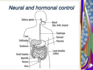
Neurotransmitter Action • Release of neurotransmitters from presynaptic vesicles requires an influx of calcium ions, Ca++ • Postsynaptic membrane receptors bind the neurotransmitter and initiate the response • Example: A neuromuscular junction and the neurotransmitter acetylcholine (ACh)
Neuromuscular junctions A An action potential propagates along a motor neuron. B The action potential reaches axon terminals that lie close to muscle fibers. axon of a motor neuron muscle fiber axon terminal muscle fiber Fig. 33-11 (a-b), p. 560
Close-up of a neuromuscular junction (a type of synapse) C Arrival of the action potential causes calcium ions (Ca++) to enter an axon terminal. one axon terminal of the presynaptic cell (motor neuron) plasma membrane of the postsynaptic cell (muscle cell) D Ca++ causes vesicles with signaling molecule (neurotransmitter) to move to the plasma membrane and release their contents by exocytosis. synaptic vesicle receptor protein in membrane of post-synaptic cell synaptic cleft (gap between pre- and postsynaptic cells) Fig. 33-11 (c-d), p. 560
Close-up of neurotransmitter receptor proteins in the plasma membrane of the postsynaptic cell binding site for neurotransmitter is vacant neurotransmitter in binding site ion crossing plasma membrane through the now-open channel channel through interior is closed F Neurotransmitter diffuses across the synaptic cleft and binds to the receptor protein. The ion channel opens, and ions flow passively into the postsynaptic cell. E When neurotransmitter is not present, the channel through the receptor protein is shut, and ions cannot flow through it. Fig. 33-11 (e-f), p. 560
Receiving the Signal • A neurotransmitter may have excitatory or inhibitory effects on a postsynaptic cell • Synaptic integration • Summation of all excitatory and inhibitory signals arriving at a postsynaptic cell at the same time • The neurotransmitter must be cleared from the synapse after the signal is transmitted
Synaptic Integration what action potential spiking would look like threshold excitatory signal integrated potential resting membrane potential inhibitory signal Fig. 33-12, p. 561
Neural Control Chapter 33 Part 2
33.6 A Smorgasbord of Signals • Different types of neurons release different neurotransmitters; Parkinson’s disease involves dopamine-secreting neurons and motor control Battling Parkinson’s disease. (a) This neurological disorder affects former heavyweight champion Muhammad Ali, actor Michael J. Fox, and about half a million other people in the United States. (b) A normal PET scan and (c) one from an affected person. Red and yellow indicate high metabolic activity in dopamine-secreting neurons. Section 2.2 explains PET scans.
The Neuropeptides • Neuromodulators • Neuropeptides made by some neurons that influence the effects of neurotransmitters • Substance P enhances pain • Enkephalins and endorphins are pain killers
33.7 Drugs Disrupts Signaling • Psychoactive drugs exert their effects by interfering with the action of neurotransmitters • Stimulants (nicotine, caffeine, cocaine, amphetamines) • Depressants (alcohol, barbiturates) • Analgesics (narcotics, ketamine, PCP) • Hallucinogens (LSD, THC)