Neural Control and the Senses
840 likes | 1.01k Vues
Neural Control and the Senses. Chapter 25. Neurons. Communication units of nervous systems Detect information about internal and external conditions Issue commands for responsive actions. stimulus (output). Types of Neurons. receptors. sensory neurons. Sensory neurons

Neural Control and the Senses
E N D
Presentation Transcript
Neural Control and the Senses Chapter 25
Neurons • Communication units of nervous systems • Detect information about internal and external conditions • Issue commands for responsive actions
stimulus (output) Types of Neurons receptors sensory neurons • Sensory neurons • Detect and relay information • Interneurons • Receive and process information • Motor neurons • Transmit signals from interneurons to effectors integrators interneurons of brain, spinal cord motor neurons effectors muscles, glands response (output)
Structure of a Neuron dendrites INPUT ZONE cell body axon OUPUT ZONE TRIGGER ZONE CONDUCTING ZONE axon endings
Neuroglia • Cells that metabolically assist, structurally support, and protect neurons • Make up more than half the volume of the vertebrate nervous system
Resting Membrane Potential • Electrical gradient across membrane • About -70 mV • Maintained by sodium-potassium pump • Potassium (K+) higher inside • Sodium (Na+) higher outside more Na+ flows into the neuron neuron becomes more positive inside more gated channels for Na+ open
Na+ K+ outside plasma membrane inside Na+ K+ p.424a
How Ions Move across Membrane interstitial fluid cytoplasm Na+/K+ pump passive transporters with open channels passive transporters with voltage-sensitive gated channels active transporters lipid bilayer of neuron membrane
Action Potential • Brief reversal in membrane potential • Voltage change causes voltage-gated channels in membrane to open • Inside of neuron briefly becomes more positive than outside
Action Potential 1 2 Na+ Na+ Na+ K+ K+ K+ K+ K+ K+ K+ Na+ Na+ Na+ Na+ 3 4 Na+ Na+
interstitial fluid cytoplasm Fig. 25-4a, p.425
Na+ Na+ Na+ Fig. 25-4b, p.425
K+ K+ K+ Na+ Na+ Na+ Fig. 25-4c, p.425
Na+/K+ pump K+ K+ K+ Na+ Na+ Na+ K+ Fig. 25-4d, p.425
Positive Feedback more Na+ ions flow into the neuron more gated channels for Na+ open neuron becomes more positive inside
All or Nothing • All action potentials are the same size • If stimulation is below threshold level, no action potential occurs • If stimulation is above threshold level, cell always depolarizes to same level
Repolarization • Once action potential peak is reached, Na+ gates close and K+ gates open • Movement of K+ out of cell • The inside of the cell once again becomes more negative than the outside
Recording of Action Potential action potential +20 0 -20 Membrane potential (millivolts) threshold -40 resting membrane potential -70 5 0 2 3 1 4 Time (milliseconds)
Propagation of Action Potentials • Action potential in one part of an axon brings neighboring region to threshold • Action potential moves from one patch of membrane to another • Can only move one direction
Chemical Synapses • Action potentials cannot jump from cell to cell • Signal is transmitted from axon end, across a synaptic cleft, by chemical signals called neurotransmitters
Chemical Synapse • Gap between the terminal ending of an axon and the input zone of another cell plasma membrane of axon ending of presynaptic cell plasma membrane of postsynaptic cell synaptic vesicle synaptic cleft membrane receptor
Synaptic Transmission • Action potential in axon ending triggers release of neurotransmitter from presynaptic cell into synaptic cleft vesicle inside presynaptic cell synaptic cleft postsynaptic cell
Synaptic Transmission • Neurotransmitter diffuses across cleft and binds to receptors on membrane of postsynaptic cell • Binding of neurotransmitter to receptors opens ion gates in membrane of postsynaptic cell
Ion Gates Open neurotransmitter ions receptor for neurotransmitter gated channel protein
Synaptic Integration • Many signals reach a neuron at the same time • Signals may suppress or reinforce one another • Whether or not an action potential occurs depends on the sum of the signals the neuron receives
Neuromuscular Junction • Synapse between motor neuron and skeletal muscle fiber • Neuron releases chemical neurotransmitter acetylcholine (ACh)
A Neuromuscular Junction neuromuscular junction motor neuron axons from spinal cord to skeletal muscle fibers transverse slice of spinal cord part of a skeletal muscle Fig. 25-6a, p.427
A Neuromuscular Junction muscle fiber axon ending Fig. 25-6b, p.427
Neurotransmitters • Acetylcholine (ACh) • Norepinephrine • Epinephrine • Dopamine • Serotonin • GABA
Cleaning Up • After neurotransmitter has acted, it is quickly removed from synaptic cleft • Molecules diffuse away, are pumped out, or broken down
interneuron motor neuron sensory neuron Information Flow
Organization • Neurons are bundled in nerves • Nerves are organized in circuits and reflex pathways • Information from sensory neurons is relayed to interneurons in spinal cord and brain • Motor neurons carry signals to body
Nerve axon myelin sheath • A bundle of axons enclosed within a connective tissue sheath many neurons inside a connective tissue sheath
Myelin Sheath • Sheath blocks ion movements • Action potential must “jump” from node to node • Greatly enhances speed of transmission
Multiple Sclerosis • A condition in which nerve fibers lose their myelin • Slows conduction • Symptoms include visual problems, numbness, muscle weakness, and fatigue
Reflexes • Automatic movements in response to stimuli • In simplest reflex arcs, sensory neurons synapse directly on motor neurons • Most reflexes involve an interneuron
Stretch Reflex STIMULUS Biceps stretches. sensoryneuron motorneuron RESPONSE Biceps contracts.
Invertebrate Nervous Systems • All animals except sponges have some sort of nervous system • Nerve cells interact with one another in signal-conducting and information-processing highways
Bilateral Nervous System rudimentary brain branching nerve nerve cord ganglion (one in most body segments)
Vertebrate Development • Earliest fishlike vertebrates had a hollow, tubular nerve cord • Modification and expansion of nerve cord produced spinal cord and brain • Nerve cord persists in vertebrate embryos as a neural tube
Central and Peripheral Nervous Systems • Central nervous system (CNS) • Brain • Spinal cord • Peripheral nervous system • Nerves that thread through the body
Major Nerves Brain cervical nerves (eight pairs) cranial nerves (twelve pairs) Spinal Cord thoracic nerves (twelve pairs) ulnar nerve (one in each arm) lumbar nerves (five pairs) sacral nerves (five pairs) sciatic nerve (one in each leg) coccygeal nerves (one pair) Fig. 25-12, p.431
Peripheral Nervous System • Somatic nerves • Motor functions • (Shown in green) • Autonomic nerves • Visceral functions • (Shown in red)
Two Types of Autonomic Nerves Sympathetic Parasympathetic • Most organs receive input from both • Usually have opposite effects on organ
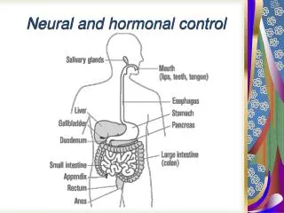
optic nerve midbrain eggs medulla oblongata salivary glands heart vagus nerve cervical nerves (8pairs) larynx bronchi lungs stomach thoracic nerves (12 pairs) liver spleen pancreas kidneys adrenal glands small intestine upper colon lower colon rectum lumbar nerves (five pairs) (all ganglia in walls of organs) (most ganglia near spinal cord) sacral nerves (five pairs) bladder uterus pelvic nerve genitals Autonomic Nervous System Fig. 25-13, p.432
Sympathetic Nerves • Originate in thoracic and lumbar regions of spinal cord • Ganglia are near the spinal cord • Respond to stress or physical activity (fight-or-flight response)
Parasympathetic Nerves • Originate in brain and sacral region of spinal cord • Ganglia are in walls of organs • Promote housekeeping responses such as digestion
Opposing Systems • Most organs receive both sympathetic and parasympathetic signals • Example: Sympathetic nerves signal heart to speed up; parasympathetic stimulate it to slow down • Synaptic integration determines response